NEET Ug . NEET Chemistry . Chemistry . Biomolecules
Chapter 19: Biomolecules
Ch 19
NEET Chemistry
NEET Ug
Carbohydrates
Definition and General Properties
Optically active polyhydroxy aldehydes or ketones, or compounds yielding these on hydrolysis.
General formula: Cx(H2O)y, considered hydrates of carbon.
Examples: glucose, sucrose, starch.
Sugars are sweet carbohydrates; sucrose (table sugar) and lactose (milk sugar) are common examples.
Also called saccharides (Greek: sakcharon = sugar).
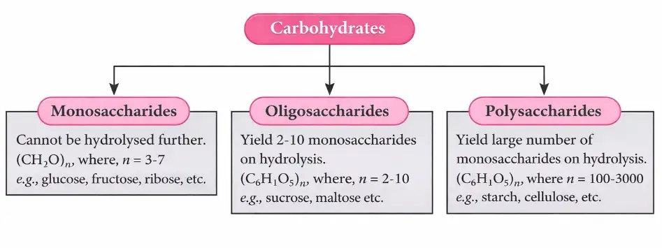
Classification Based on Hydrolysis
Monosaccharides: Simple sugars, cannot be hydrolyzed further (e.g., glucose, fructose).
Oligosaccharides: 2-10 monosaccharide units; hydrolyze to monosaccharides (e.g., sucrose, maltose).
Polysaccharides: Long chains of monosaccharides (100+ units); hydrolyze to many monosaccharides (e.g., starch, cellulose).
Classification Based on Reducing Properties
Reducing sugars: Contain free aldehyde or ketone groups; reduce Fehling's and Tollens' reagents (e.g., glucose, maltose, lactose).
Non-reducing sugars: No free aldehyde or ketone groups; do not reduce Fehling's or Tollens' reagents (e.g., sucrose).
Solved Examples
Ex 1: Identify if maltose is a reducing sugar.
- Maltose has a free aldehyde group at C-1 of one glucose unit.
- It reduces Fehling's solution.
- Therefore, maltose is a reducing sugar.
Ex 2: Hydrolysis of sucrose.
- Sucrose + H2O (acid/enzyme) → glucose + fructose.
- Both monosaccharides are reducing sugars, but sucrose itself is non-reducing.
Practice Set
Q1: Which of the following is a non-reducing sugar?
A. Glucose
B. Maltose
C. Sucrose
D. LactoseQ2: Oligosaccharides contain how many monosaccharide units?
A. 1
B. 2-10
C. 10-100
D. More than 100Q3: Which reagent is used to test reducing sugars?
A. Benedict's solution
B. Fehling's solution
C. Tollens' reagent
D. All of the above
Answer Key
Q1: C. Sucrose - It lacks free aldehyde or ketone groups and does not reduce Fehling's or Tollens' reagents.
Q2: B. 2-10 - Oligosaccharides consist of 2 to 10 monosaccharide units.
Q3: D. All of the above - Benedict's, Fehling's, and Tollens' reagents all test for reducing sugars.
Memory Aids
"Mono" = single sugar unit; "Oligo" = few units; "Poly" = many units.
Reducing sugars have free aldehyde/ketone groups; non-reducing sugars do not.
Sucrose is non-reducing despite being a disaccharide.
Monosaccharides
Classification and Properties
Classified by carbon number: trioses (3C), pentoses (5C), hexoses (6C), etc.
Based on functional group: Aldoses (aldehyde group), Ketoses (ketone group).
Glucose: aldohexose (6C, aldehyde), naturally occurring, found in fruits and honey.
Fructose: ketohexose (6C, ketone), found in fruits and honey, sweeter than glucose.
Preparation of Glucose
From sucrose: Boiling sucrose with dilute HCl or H2SO4 in alcoholic solution yields equal glucose and fructose.
From starch: Hydrolysis of starch with dilute H2SO4 at 393 K under pressure produces glucose.


Structure of Glucose
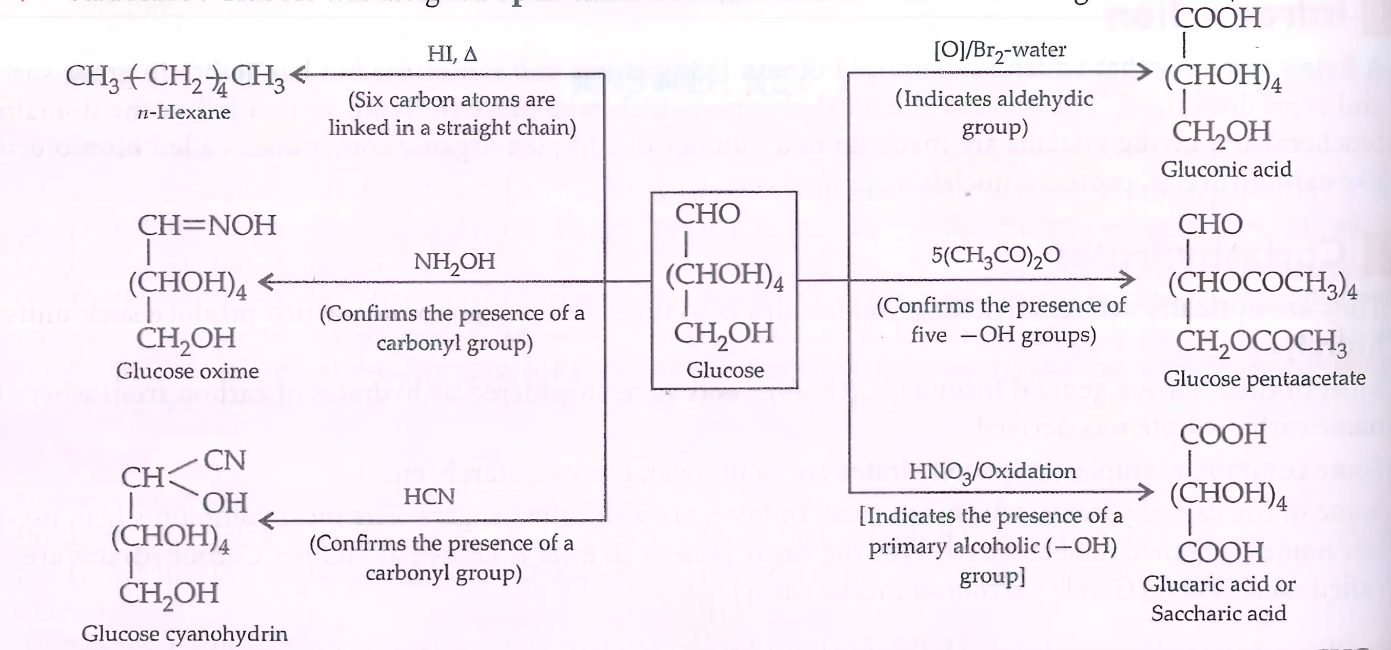
Open chain form confirmed by reactions with HI, NH2OH, HCN, bromine water.
>
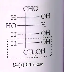
Fischer gave exact spatial arrangement of —OH groups.
Glucose exists as cyclic hemiacetal (pyranose) forms: α and β anomers differ at C-1 (anomeric carbon).
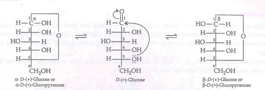
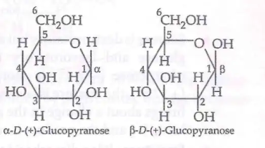
Haworth projection represents cyclic glucose.
Other Reactions of Glucose
Reduction: Glucose → D-Sorbitol (using Na-Hg).
Oxidation: Glucose → Gluconic acid (Fehling's or Tollens' test positive).
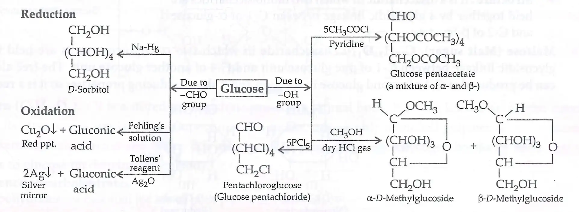
Derivatives: Glucose pentaacetate (acetic anhydride), pentachloroglucose (PCl5), methylglucosides (CH3OH + HCl).
Solved Examples
Ex 1: Hydrolysis of sucrose.
- Sucrose + H2O (acid) → glucose + fructose (1:1 ratio).
Ex 2: Identify the anomeric carbon in glucose.
- Carbon 1 (C-1) is the anomeric carbon where α and β forms differ.
Practice Set
Q1: Glucose is classified as:
A. Ketohexose
B. Aldohexose
C. Aldopentose
D. KetopentoseQ2: Which reagent confirms the aldehyde group in glucose?
A. HI and heat
B. NH2OH
C. Bromine water
D. All of the aboveQ3: The cyclic form of glucose is called:
A. Furanose
B. Pyranose
C. Glucopyranose
D. Both B and C
Answer Key
Q1: B. Aldohexose - Glucose has six carbons and an aldehyde group.
Q2: D. All of the above - These reagents confirm the aldehyde group in glucose.
Q3: D. Both B and C - The six-membered cyclic form is pyranose, also called glucopyranose.
Memory Aids
Glucose = aldohexose; fructose = ketohexose.
Anomeric carbon = C-1 in glucose; α and β differ in OH position here.
Pyranose = six-membered ring; furanose = five-membered ring.
Fructose
Structure and Properties
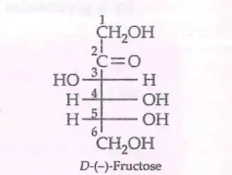
Ketohexose: 6 carbons, ketone group at C-2.
Found in fruits, honey, vegetables; used as sweetener.
Belongs to D-series; laevorotatory (D-(-)-fructose).
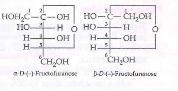
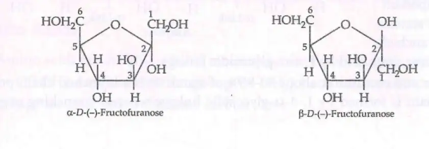
Exists in two cyclic forms (furanose rings) formed by OH at C-5 attacking ketone at C-2.
α and β anomers differ in OH position at C-2.
Haworth Structures and Isomerism
Cyclic forms represented by Haworth projections.
Fructose anomers: α-D-(−)-Fructofuranose and β-D-(−)-Fructofuranose.
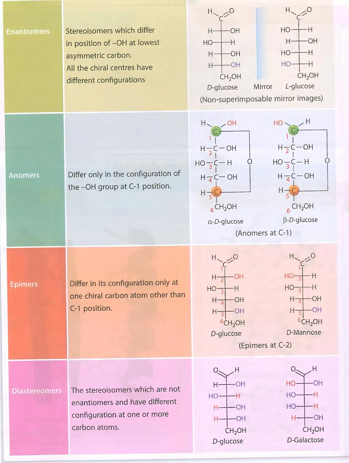
Isomer types: Enantiomers, Anomers, Epimers, Diastereomers (differences in OH group positions).
Solved Examples
Ex 1: Identify the ring size of fructose cyclic form.
- Fructose forms a five-membered furanose ring.
Ex 2: Difference between α and β fructofuranose.
- Position of OH on C-2: down in α, up in β.
Practice Set
Q1: Fructose is classified as:
A. Aldohexose
B. Ketohexose
C. Aldopentose
D. KetopentoseQ2: The cyclic form of fructose is:
A. Pyranose
B. Furanose
C. Glucopyranose
D. None of the aboveQ3: The difference between α and β fructofuranose is:
A. Position of OH on C-1
B. Position of OH on C-2
C. Number of carbons
D. Type of sugar
Answer Key
Q1: B. Ketohexose - Fructose has a ketone group at C-2.
Q2: B. Furanose - Fructose forms a five-membered ring.
Q3: B. Position of OH on C-2 - α and β differ at C-2 OH position.
Memory Aids
Fructose = ketohexose with furanose ring.
α and β anomers differ at C-2 OH position.
Disaccharides
Structure and Properties

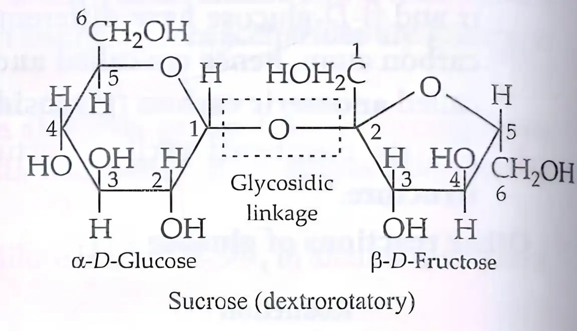
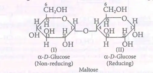
Formed by two monosaccharides linked via glycosidic (oxide) linkage.
Glycosidic bond formed by loss of water between OH groups.
Examples:
Sucrose: Glucose + Fructose; non-reducing sugar; hydrolyzes to invert sugar.
Maltose: Two α-D-glucose units; reducing sugar.
Lactose: β-D-galactose + β-D-glucose; reducing sugar.
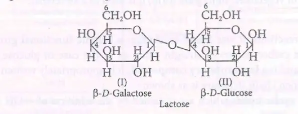
Hydrolysis Reactions
Sucrose + H2O (invertase or acid) → glucose + fructose (invert sugar).
Maltose + H2O (maltase) → 2 glucose molecules.
Lactose + H2O (lactase) → galactose + glucose.
Solved Examples
Ex 1: Hydrolysis of sucrose.
- Sucrose + H2O → glucose + fructose (invert sugar).
Ex 2: Identify reducing sugar among disaccharides.
- Maltose and lactose are reducing sugars; sucrose is non-reducing.
Practice Set
Q1: Sucrose is a disaccharide of:
A. Glucose + Glucose
B. Glucose + Fructose
C. Galactose + Glucose
D. Fructose + GalactoseQ2: Which disaccharide is non-reducing?
A. Maltose
B. Lactose
C. Sucrose
D. CellobioseQ3: Glycosidic linkage in maltose is between:
A. C-1 and C-4 of two glucose units
B. C-1 and C-6 of two glucose units
C. C-1 and C-4 of glucose and fructose
D. C-1 and C-4 of galactose and glucose
Answer Key
Q1: B. Glucose + Fructose - Sucrose is composed of these monosaccharides.
Q2: C. Sucrose - It lacks free aldehyde/ketone groups.
Q3: A. C-1 and C-4 of two glucose units - Maltose linkage is α-1,4 glycosidic bond.
Memory Aids
Sucrose = glucose + fructose, non-reducing.
Maltose = two glucose units, reducing sugar.
Lactose = galactose + glucose, reducing sugar.
Polysaccharides
Types and Structure
Starch: Plant storage polysaccharide; polymer of α-D-glucose.
Components:
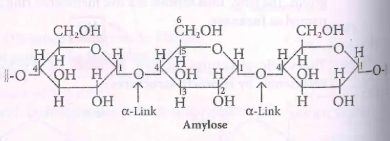
Amylose: linear, 200-1000 α-D-glucose units linked by α-1,4 glycosidic bonds; water soluble.
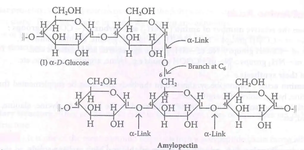
Amylopectin: branched polymer with α-1,4 linkages and α-1,6 branches at C-6.
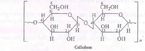
Cellulose: Structural polysaccharide in plants; linear polymer of β-D-glucose linked by β-1,4 glycosidic bonds.
Glycogen: Animal storage polysaccharide; highly branched α-D-glucose polymer similar to amylopectin; stored in liver, muscles, brain.
Properties and Importance
Cellulose forms plant cell walls; provides strength and rigidity.
Starch and glycogen serve as energy storage in plants and animals respectively.
Glycogen is broken down enzymatically to glucose on demand.
Cellulose is used in cotton fibers, wood, and bacterial cell walls.
Mercerisation: treatment of cellulose fibers with NaOH to impart lustre.
Solved Examples
Ex 1: Identify the glycosidic linkage in cellulose.
- β-1,4 glycosidic bonds between β-D-glucose units.
Ex 2: Difference between amylose and amylopectin.
- Amylose: linear α-1,4 linkages.
- Amylopectin: branched with α-1,4 and α-1,6 linkages.
Practice Set
Q1: The main storage polysaccharide in plants is:
A. Cellulose
B. Glycogen
C. Starch
D. DextranQ2: Glycogen is stored mainly in:
A. Liver and muscles
B. Brain and bones
C. Kidneys
D. LungsQ3: The glycosidic linkage in cellulose is:
A. α-1,4
B. β-1,4
C. α-1,6
D. β-1,6
Answer Key
Q1: C. Starch - Main plant storage polysaccharide.
Q2: A. Liver and muscles - Glycogen storage sites.
Q3: B. β-1,4 - Cellulose linkage type.
Memory Aids
Starch = plant storage; glycogen = animal storage.
Amylose = linear; amylopectin = branched.
Cellulose = β-1,4 linkages; indigestible by humans.
Importance of Carbohydrates
Biological Roles
Primary energy source for plants and animals.
Storage molecules: starch in plants, glycogen in animals.
Structural components: cellulose in plant cell walls, bacterial cell walls.
Constituents of nucleic acids: D-ribose and 2-deoxy-D-ribose sugars.
Natural sugars mostly exist in D-form.
Bacterial enzyme converts sucrose to dextran, a polysaccharide.
Mercerisation of cotton (cellulose) with NaOH improves fabric lustre.
Solved Examples
Ex 1: Name the sugar in nucleic acids.
- D-ribose and 2-deoxy-D-ribose.
Ex 2: What is mercerisation?
- Treatment of cellulose fibers with concentrated NaOH to enhance lustre.
Practice Set
Q1: The sugar present in nucleic acids is:
A. Glucose
B. D-ribose
C. Fructose
D. LactoseQ2: Mercerisation is a treatment of:
A. Starch
B. Glycogen
C. Cellulose
D. DextranQ3: Dextran is a polysaccharide produced by:
A. Plants
B. Animals
C. Bacteria
D. Fungi
Answer Key
Q1: B. D-ribose - Sugar in nucleic acids.
Q2: C. Cellulose - Mercerisation treats cellulose fibers.
Q3: C. Bacteria - Bacterial enzyme converts sucrose to dextran.
Memory Aids
Nucleic acids contain ribose sugars.
Mercerisation = NaOH treatment of cotton (cellulose).
Dextran produced by bacteria from sucrose.
Proteins
Overview
Proteins are polymers of α-amino acids linked by peptide bonds.
Essential for structure, function, growth, and maintenance of the body.
Main sources: milk, cheese, pulses, peanuts, fish, meat.
Solved Examples
Ex 1: Identify the monomer of proteins.
Answer: α-amino acids.
Practice Set
Q1: Proteins are polymers of:
A. Monosaccharides
B. Fatty acids
C. α-amino acids
D. NucleotidesQ2: Which of the following is a main source of protein?
A. Glucose
B. Milk
C. Starch
D. CelluloseQ3: Proteins are important for:
A. Energy storage
B. Growth and maintenance
C. Photosynthesis
D. Respiration
Answer Key
Q1: C - Proteins are made of α-amino acids.
Q2: B - Milk is a rich protein source.
Q3: B - Proteins are essential for growth and maintenance.
Memory Aids
Protein = Primary importance (Greek origin).
Protein monomer = α-amino acid.
Sources: Milk, Meat, Pulses (MMP).
Amino Acids
Structure and Properties
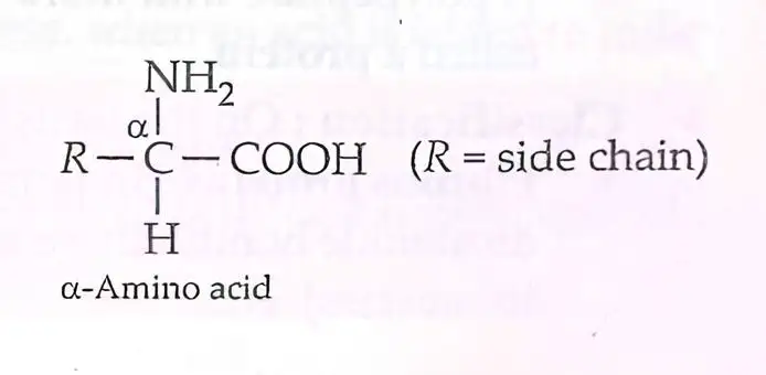
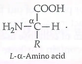
Bifunctional molecules with acidic (—COOH) and basic (—NH2) groups attached to the same α-carbon.
General formula: NH2–CH(R)–COOH, where R is side chain.
Usually colorless, crystalline, water-soluble, high melting solids.
Behave like salts due to acidic and basic groups.
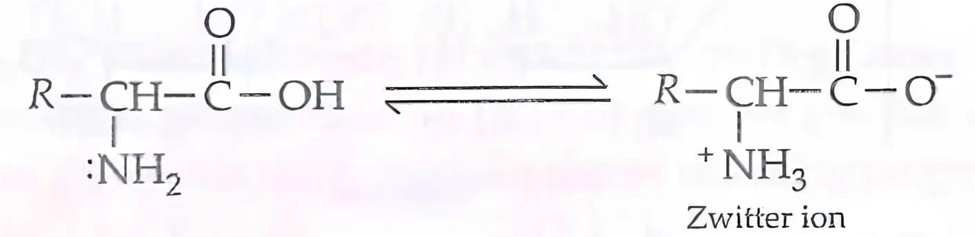
Exist as zwitterions in aqueous solution (dipolar ions with + and − charges).
Solved Examples
Ex 1: Identify the functional groups in amino acids.
Answer: Amino (—NH2) and carboxyl (—COOH).
Practice Set
Q1: Amino acids contain which functional groups?
A. Hydroxyl and aldehyde
B. Amino and carboxyl
C. Ketone and ester
D. Ether and amideQ2: Zwitterion form of amino acid has:
A. Only positive charge
B. Only negative charge
C. Both positive and negative charges
D. No chargeQ3: Amino acids are soluble in water because:
A. They are nonpolar
B. They form salts
C. They are hydrocarbons
D. They are gases
Answer Key
Q1: B - Amino acids have amino and carboxyl groups.
Q2: C - Zwitterions carry both positive and negative charges.
Q3: B - Amino acids behave like salts, increasing solubility.
Memory Aids
Remember: Amino + Acid = Amino acid.
Zwitterion = Both charges present.
Water solubility due to salt-like nature.
Classification of Amino Acids
Types Based on Functional Groups
Neutral: Equal number of —COOH and —NH2 groups (e.g., glycine, alanine).
Acidic: More —COOH groups than —NH2 (e.g., aspartic acid, glutamic acid).
Basic: More —NH2 groups than —COOH (e.g., lysine, arginine).
Types Based on Synthesis
Essential: Cannot be synthesized by body; must be obtained from diet (e.g., valine, leucine).
Non-essential: Synthesized by body (e.g., glycine, alanine).
Solved Examples
Ex 1: Classify lysine.
Answer: Basic amino acid.Ex 2: Is valine essential or non-essential?
Answer: Essential amino acid.
Practice Set
Q1: Which amino acid is acidic?
A. Glycine
B. Aspartic acid
C. Lysine
D. AlanineQ2: Essential amino acids are:
A. Synthesized in body
B. Obtained from diet
C. Nonpolar
D. Acidic onlyQ3: Basic amino acid example is:
A. Glutamic acid
B. Arginine
C. Valine
D. Serine
Answer Key
Q1: B - Aspartic acid is acidic.
Q2: B - Essential amino acids must be obtained from diet.
Q3: B - Arginine is basic.
Memory Aids
Neutral = equal acidic and basic groups.
Essential = Eat (must be in diet).
Basic amino acids have extra amino groups.
Isoelectric Point
Concept
pH at which amino acid exists as zwitterion with equal positive and negative charges.
At this pH, amino acid does not migrate in electric field.
Solubility in water is minimum at isoelectric point.
Useful for amino acid separation.
Solved Examples
Ex 1: What happens to amino acid at isoelectric point?
Answer: Exists as neutral zwitterion, no migration in electric field.
Practice Set
Q1: Isoelectric point is the pH where amino acid:
A. Is fully protonated
B. Has no net charge
C. Is fully deprotonated
D. Is insolubleQ2: At isoelectric point, amino acid:
A. Migrates to cathode
B. Migrates to anode
C. Does not migrate
D. DecomposesQ3: Amino acid solubility at isoelectric point is:
A. Maximum
B. Minimum
C. Zero
D. Unchanged
Answer Key
Q1: B - Net charge is zero at isoelectric point.
Q2: C - No migration due to neutral charge.
Q3: B - Solubility is minimum at isoelectric point.
Memory Aids
Isoelectric = "iso" means equal charges.
Neutral charge = no movement in electric field.
Solubility dips at isoelectric point.
Peptide Bond
Formation and Structure
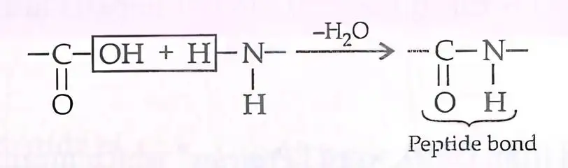
Peptide bond is an amide bond formed between —COOH of one amino acid and —NH2 of another.
Formed by condensation reaction with elimination of H2O.
Links amino acids to form peptides and proteins.
Solved Examples
Ex 1: Write the reaction for peptide bond formation.
Answer: Amino acid 1 (—COOH) + Amino acid 2 (—NH2) → Peptide bond + H2O
Practice Set
Q1: Peptide bond is formed between:
A. Two amino groups
B. Two carboxyl groups
C. Amino and carboxyl groups
D. Side chainsQ2: Peptide bond formation involves:
A. Hydrolysis
B. Condensation
C. Oxidation
D. ReductionQ3: Peptides with 2-10 amino acids are called:
A. Polypeptides
B. Oligopeptides
C. Proteins
D. Monopeptides
Answer Key
Q1: C - Peptide bond forms between amino and carboxyl groups.
Q2: B - Condensation reaction with water loss.
Q3: B - Oligopeptides contain 2-10 amino acids.
Memory Aids
Peptide bond = Amide bond.
Condensation = Water removed.
Oligo = Few (2-10 amino acids).
Protein Structure
Levels of Structure
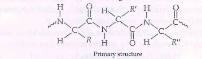
Primary: Linear sequence of amino acids linked by peptide bonds.
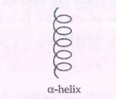
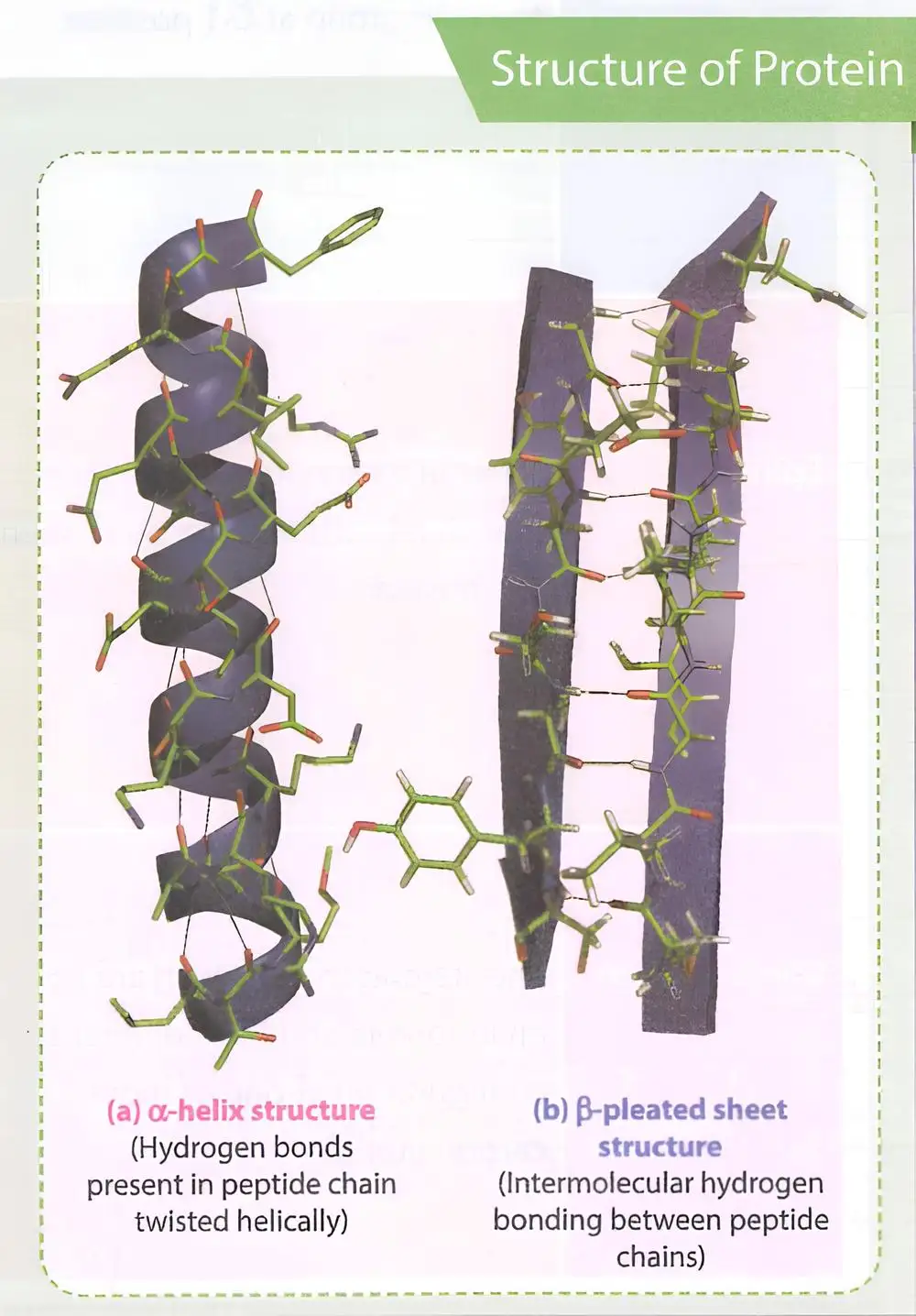
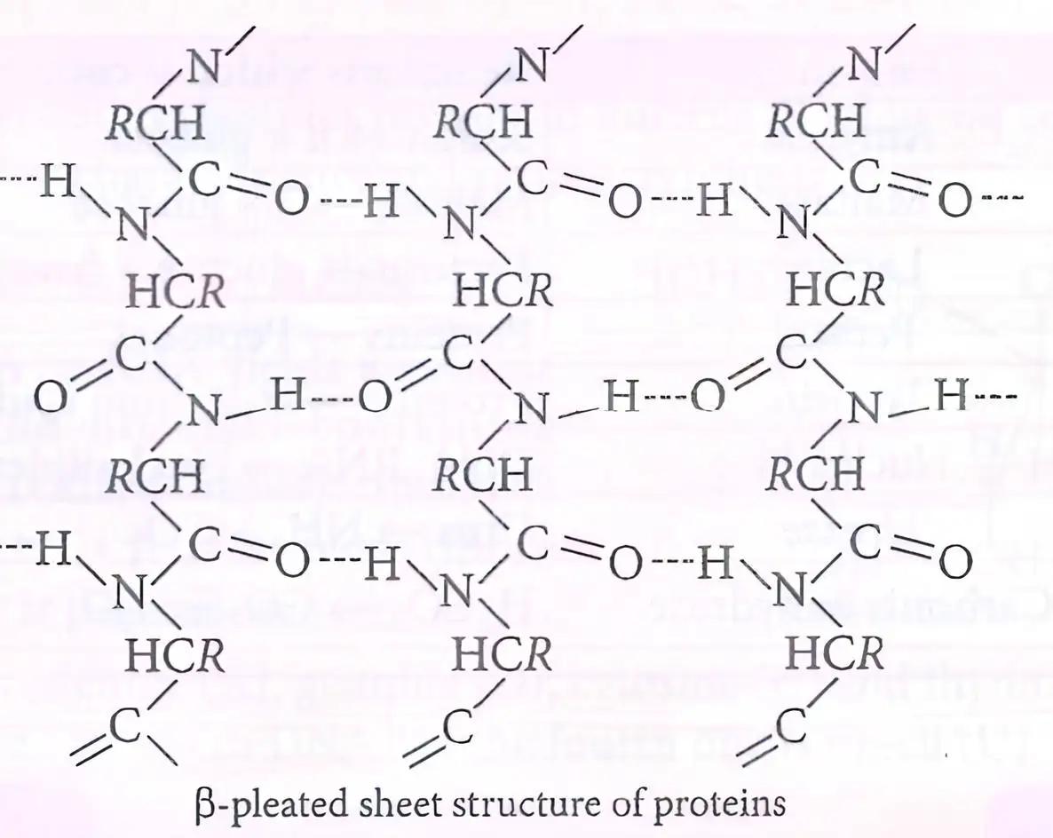
Secondary: α-helix and β-pleated sheet formed by hydrogen bonding.
Tertiary: Overall 3D folding stabilized by hydrogen bonds, disulfide bridges, van der Waals, electrostatic forces.
Quaternary: Arrangement of multiple polypeptide subunits.
Solved Examples
Ex 1: Name two secondary structures.
Answer: α-helix and β-pleated sheet.Ex 2: What stabilizes tertiary structure?
Answer: Hydrogen bonds, disulfide linkages, van der Waals, electrostatic forces.
Practice Set
Q1: Primary structure of protein is:
A. 3D folding
B. Sequence of amino acids
C. Multiple subunits
D. Hydrogen bonding patternQ2: α-helix is stabilized by:
A. Ionic bonds
B. Hydrogen bonds
C. Covalent bonds
D. Metallic bondsQ3: Quaternary structure involves:
A. Single polypeptide chain
B. Multiple polypeptide subunits
C. Peptide bond formation
D. Amino acid sequence
Answer Key
Q1: B - Primary structure is amino acid sequence.
Q2: B - α-helix stabilized by hydrogen bonds.
Q3: B - Quaternary structure is arrangement of subunits.
Memory Aids
Primary = Sequence.
Secondary = α-helix & β-sheet.
Tertiary = 3D folding.
Quaternary = Multiple subunits.
Denaturation of Proteins
Concept
Loss of biological activity due to disruption of secondary and tertiary structures.
Caused by physical (temperature) or chemical (pH) changes.
Primary structure remains intact.
Examples: Coagulation of egg white on boiling, curdling of milk.
Solved Examples
Ex 1: What happens to protein on boiling?
Answer: Denaturation; loss of structure and function.
Practice Set
Q1: Denaturation affects which protein structure?
A. Primary
B. Secondary and tertiary
C. Quaternary only
D. NoneQ2: Denaturation can be caused by:
A. Change in temperature
B. Change in pH
C. Both A and B
D. NoneQ3: Which remains unchanged during denaturation?
A. Primary structure
B. Secondary structure
C. Tertiary structure
D. Biological activity
Answer Key
Q1: B - Secondary and tertiary structures are affected.
Q2: C - Both temperature and pH changes cause denaturation.
Q3: A - Primary structure remains intact.
Memory Aids
Denaturation = Unfolding, loss of function.
Primary structure = Peptide bonds intact.
Heat and pH = Common denaturants.
Enzymes
Overview
Biological catalysts that speed up biochemical reactions.
Mostly globular proteins with high specificity for substrates and products.
Named after the substrate or reaction they catalyze (e.g., maltase hydrolyzes maltose).
Increase reaction rates significantly without being consumed.

Solved Examples
Ex 1: Hydrolysis of maltose by maltase
Maltose + H2O → 2 Glucose (catalyzed by maltase)
Water breaks the glycosidic bond with enzyme assistance.
Practice Set
Q1: What type of biomolecule are most enzymes?
A. Lipids
B. Carbohydrates
C. Globular proteins
D. Nucleic acids
Q2: The enzyme maltase catalyzes the breakdown of:
A. Glucose into maltose
B. Maltose into glucose
C. Lactose into glucose and galactose
D. Sucrose into fructose and glucose
Q3: Which statement about enzymes is correct?
A. They are consumed in the reaction.
B. They decrease reaction specificity.
C. They increase activation energy.
D. They speed up reactions without being consumed.
Answer Key
Q1: C - Enzymes are mostly globular proteins.
Q2: B - Maltase hydrolyzes maltose into glucose.
Q3: D - Enzymes speed up reactions and are not consumed.
Memory Aids
Enzymes = Globular proteins acting as biological catalysts.
"-ase" suffix indicates enzyme name related to substrate.
Hydrolysis reactions often involve water and enzymes like maltase.
Mechanism of Enzyme Action
Key Points
Enzymes lower activation energy, speeding up reactions.
Only small amounts of enzyme are needed for reaction progress.
Example: Activation energy for acid hydrolysis of sucrose is 6.22 kJ mol−1, reduced to 2.15 kJ mol−1 by sucrase.
Enzyme deficiency can cause diseases (e.g., phenylalanine hydroxylase deficiency causes phenylketonuria).
Tyrosinase deficiency leads to albinism (reduced melanin pigment).
Solved Examples
Ex 1: Activation energy reduction by enzyme
Without enzyme: 6.22 kJ mol−1
With sucrase: 2.15 kJ mol−1
Enzyme lowers energy barrier, increasing reaction rate.
Practice Set
Q1: What is the main effect of enzymes on activation energy?
A. Increase it
B. Decrease it
C. No effect
D. Eliminate it
Q2: Phenylketonuria is caused by deficiency of which enzyme?
A. Tyrosinase
B. Phenylalanine hydroxylase
C. Sucrase
D. Maltase
Q3: Which condition results from tyrosinase deficiency?
A. Phenylketonuria
B. Albinism
C. Diabetes
D. Scurvy
Answer Key
Q1: B - Enzymes decrease activation energy.
Q2: B - Phenylketonuria is due to phenylalanine hydroxylase deficiency.
Q3: B - Tyrosinase deficiency causes albinism.
Memory Aids
Enzymes lower activation energy, not consumed.
Phenylketonuria = Phenylalanine hydroxylase deficiency.
Albinism = Tyrosinase deficiency (melanin pigment loss).
Common Enzymes
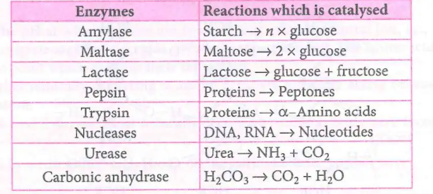
Important Enzymes and Reactions
Amylase: Starch → Glucose
Maltase: Maltose → 2 Glucose
Lactase: Lactose → Glucose + Fructose
Pepsin & Trypsin: Proteins → Peptides
Nucleases: DNA/RNA → Nucleotides
Urease: Urea → NH3 + CO2
Carbonic anhydrase: H2CO3 → CO2 + H2O
Solved Examples
Ex 1: Lactase action
Lactose + H2O → Glucose + Fructose (catalyzed by lactase)
Ex 2: Urease reaction
Urea + H2O → NH3 + CO2 (catalyzed by urease)
Practice Set
Q1: Which enzyme breaks down starch into glucose?
A. Maltase
B. Amylase
C. Lactase
D. Pepsin
Q2: Carbonic anhydrase catalyzes the conversion of:
A. CO2 to H2CO3
B. H2CO3 to CO2 and H2O
C. Urea to NH3 and CO2
D. Proteins to peptides
Q3: Which enzyme breaks down proteins into peptides?
A. Trypsin
B. Nuclease
C. Lactase
D. Urease
Answer Key
Q1: B - Amylase breaks starch into glucose.
Q2: B - Carbonic anhydrase converts H2CO3 to CO2 and H2O.
Q3: A - Trypsin breaks proteins into peptides.
Memory Aids
Amylase = starch; Maltase = maltose; Lactase = lactose.
Pepsin & Trypsin = protein digestion.
Urease = urea to ammonia and CO2.
Carbonic anhydrase = carbonic acid to CO2 and water.
Vitamins
Definition and Importance
Organic compounds needed in small amounts in diet.
Essential for specific biological functions and normal growth.
Cannot be synthesized sufficiently by the body.
Classification of Vitamins
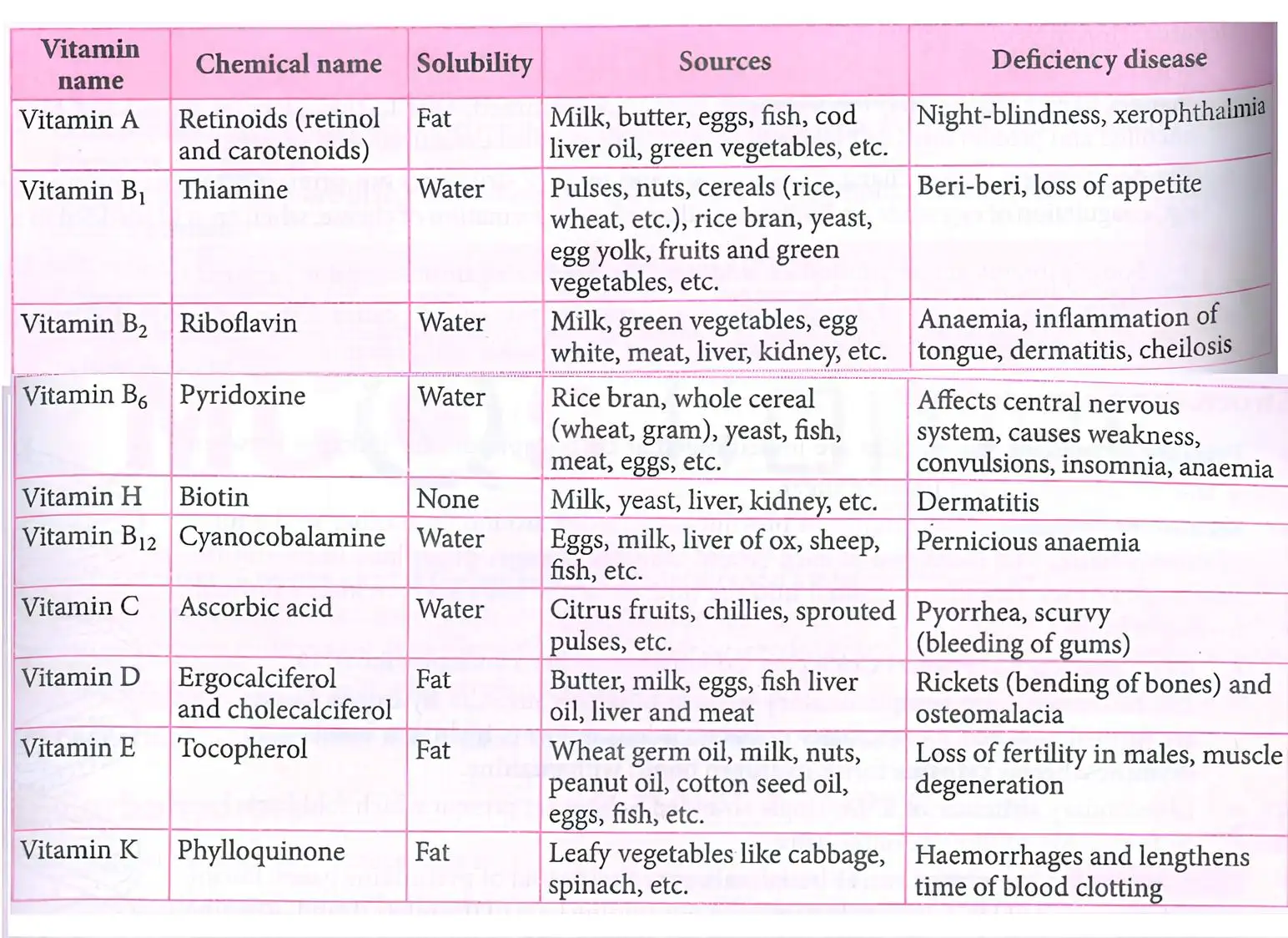
Water soluble vitamins: Dissolve in water; not stored; require regular intake; excreted in urine (except B12).
Examples: Vitamin B1 (Thiamine), B2 (Riboflavin), B6 (Pyridoxine), B12 (Cobalamin), C (Ascorbic acid).
Fat soluble vitamins: Dissolve in fats/oils; stored in liver and adipose tissue.
Examples: Vitamins A (Retinoids), D, E, K.
Key Facts
Vitamin deficiency causes avitaminosis.
Excess fat soluble vitamins cause hypervitaminosis.
Water soluble vitamins cannot be stored in the body.
Vitamin H (Biotin) is neither water nor fat soluble.
Solved Examples
Ex 1: Identify the vitamin deficient in night blindness.
Solution: Vitamin A deficiency causes night blindness.Ex 2: Which vitamin deficiency causes scurvy?
Solution: Vitamin C deficiency causes scurvy.
Practice Set
Q1: Which vitamin is stored in the liver and adipose tissue?
A. Vitamin B1
B. Vitamin C
C. Vitamin A
D. Vitamin B12Q2: Which vitamin deficiency leads to pernicious anemia?
A. Vitamin B12
B. Vitamin D
C. Vitamin K
D. Vitamin EQ3: Excess intake of which vitamin can cause hypervitaminosis?
A. Vitamin C
B. Vitamin B6
C. Vitamin A
D. Vitamin B1
Answer Key
Q1: C. Vitamin A - It is fat soluble and stored in liver and adipose tissue.
Q2: A. Vitamin B12 - Deficiency causes pernicious anemia.
Q3: C. Vitamin A - Excess fat soluble vitamins like A cause hypervitaminosis.
Memory Aids
Water soluble vitamins: B-complex and C; need regular intake.
Fat soluble vitamins: A, D, E, K; stored in body fat.
Avitaminosis = vitamin deficiency diseases.
Hypervitaminosis = excess fat soluble vitamin toxicity.
Nucleic Acids
Chemical Composition
Nucleic acids are polymers of nucleotides found in the nucleus of all living cells.
Complete hydrolysis yields pentose sugar, phosphoric acid, and nitrogenous bases.
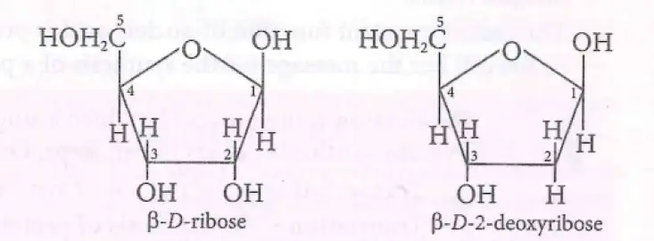
DNA contains β-deoxyribose; RNA contains β-D-ribose.
DNA bases: Adenine (A), Guanine (G), Cytosine (C), Thymine (T).
RNA bases: Adenine (A), Guanine (G), Cytosine (C), Uracil (U) replaces thymine.
Nucleosides and Nucleotides
Nucleoside = Pentose sugar + nitrogenous base.
Nucleotide = Nucleoside + phosphate group.
Nucleotides are building blocks of DNA and RNA.
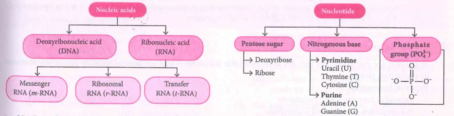
Phosphate group has one phosphorus atom bonded to four oxygen atoms, carrying a negative charge.
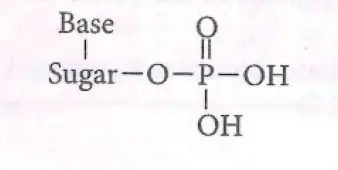
Structure of Nucleic Acids
Primary structure: Nucleotides linked by phosphodiester bonds between 5' and 3' carbons of sugar.
Secondary structure: Two polynucleotide strands coil into a double helix.
Backbone: Sugar-phosphate units; bases face inward and pair via hydrogen bonds.
Watson and Crick proposed the double helix model for DNA.
Base pairing: Adenine pairs with Thymine (2 H-bonds), Guanine pairs with Cytosine (3 H-bonds).
RNA secondary structure: Single strand folds to form double helices.
Chargaff's rule: Purine = Pyrimidine amount; A = T, G = C in DNA.
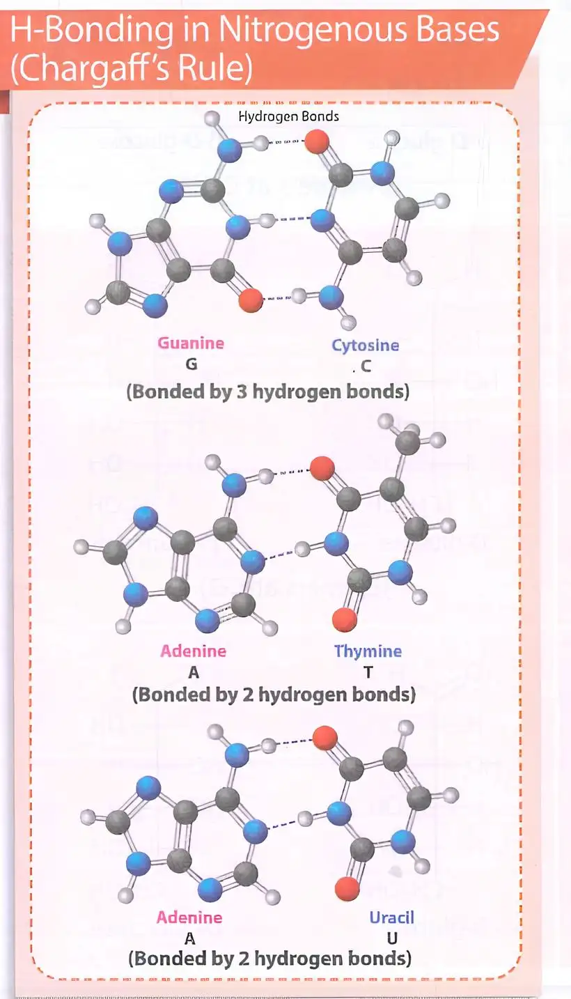
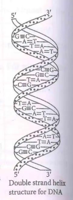
Biological Functions
DNA stores genetic information and transmits heredity.
Maintains species identity.
Self-duplicates during cell division (replication).
RNA synthesizes proteins using DNA's instructions.
Protein synthesis involves transcription (RNA synthesis) and translation (protein assembly).
Genetic code: Triplet nucleotide sequences determine amino acid sequence in proteins.
Solved Examples
Ex 1
Identify sugar in DNA and RNA: DNA has deoxyribose; RNA has ribose.
Explain base pairing: A pairs with T (DNA) or U (RNA), G pairs with C.
Practice Set
Q1: Which sugar is present in RNA?
A. β-D-deoxyribose
B. β-D-ribose
C. Glucose
D. Fructose
Q2: How many hydrogen bonds form between guanine and cytosine?
A. One
B. Two
C. Three
D. Four
Q3: Which component is NOT part of a nucleotide?
A. Phosphate group
B. Nitrogenous base
C. Pentose sugar
D. Amino acid
Answer Key
Q1: B. β-D-ribose - RNA contains ribose sugar.
Q2: C. Three - G and C pair via three hydrogen bonds.
Q3: D. Amino acid - Not a nucleotide component.
Memory Aids
RNA has ribose; DNA has deoxyribose (missing oxygen at 2' carbon).
A pairs with T (or U in RNA), G pairs with C.
Purine bases (A, G) pair with pyrimidines (T/U, C).
Nucleotide = sugar + base + phosphate.
Hormones
Types of Hormones
Steroids: Estrogens and androgens; lipid-soluble molecules derived from cholesterol.
Polypeptides: Insulin and endorphins; chains of amino acids, water-soluble.
Amino Acid Derivatives: Epinephrine and norepinephrine; modified single amino acids.
Functions of Hormones
Act as intercellular messengers secreted directly into the bloodstream by endocrine glands.
Maintain balance of biological activities in the body.
Growth hormones and sex hormones regulate growth and development.
Insulin lowers blood glucose; glucagon raises blood glucose; together regulate glucose homeostasis.
Epinephrine and norepinephrine mediate responses to external stimuli (fight or flight response).
Solved Examples
Ex 1: Identify the hormone type: Insulin is a polypeptide hormone because it is a chain of amino acids.
Ex 2: Explain glucose regulation: Insulin decreases blood glucose by promoting uptake; glucagon increases glucose by stimulating glycogen breakdown.
Practice Set
Q1: Which hormone is a steroid?
A. Insulin
B. Estrogen
C. Epinephrine
D. Endorphin
Q2: Which hormone increases blood glucose level?
A. Insulin
B. Glucagon
C. Estrogen
D. Endorphin
Q3: Epinephrine and norepinephrine are derived from:
A. Cholesterol
B. Polypeptides
C. Amino acids
D. Fatty acids
Answer Key
Q1: B. Estrogen — It is a steroid hormone derived from cholesterol.
Q2: B. Glucagon — It raises blood glucose by stimulating glycogen breakdown.
Q3: C. Amino acids — Epinephrine and norepinephrine are amino acid derivatives.
Memory Aids
Steroid hormones end with '-one' or '-ol' (e.g., estrogen, androgen).
Polypeptides include insulin; remember "Insulin is a Protein".
Amino acid derivatives include epinephrine; think "Epi = amino acid derivative".
Insulin lowers glucose; glucagon raises glucose — opposite actions maintain balance.
NEET Chemistry — ALL CHAPTERS
1
Some Basic Concepts of Chemistry
2
Structure of Atom
3
Classification of Elements and Periodicity in Properties
4
Chemical Bonding and Molecular Structure
5
Thermodynamics
6
Equilibrium
7
Redox Reactions
8
Organic Chemistry - Some Basic Principles and Techniques
9
Hydrocarbons
10
Solutions
11
Electrochemistry
12
Chemical Kinetics
13
The d- and f- Block Elements
14
Coordination Compounds
15
Haloalkanes and Haloarenes
16
Alcohols, Phenols and Ethers
17
Aldehydes, Ketones and Carboxylic Acids
18
Amines
19
Biomolecules
20
The p-Block Elements (Group 13 and 14)
21
The p-Block Elements (Group 15 to 18)
22
Principles Related to Practical Chemistry