NEET Ug . NEET Zoology . Zoology . Structural Organisation in Animals
Chapter 1: Structural Organisation in Animals
Ch 1
NEET Zoology
NEET Ug
Animal Tssues
Tissues: definition and main types
Tissue — A tissue is a group of similar cells, together with intercellular substances, that share an origin and perform a specific function.
- Unicellular organisms: A single cell performs functions such as digestion, respiration, excretion and reproduction.
- Multicellular animals: Different groups of cells carry out specialised functions; tissues are organised in patterns to form organs.
- Four basic animal tissues: Epithelial, connective, muscular and neural.
| Type | Origin | Main functions |
|---|---|---|
| Epithelial | Ectoderm, endoderm and mesoderm | Protection, secretion, absorption, excretion and reproduction |
| Connective | Mesoderm | Attachment, support, storage, protection and transport |
| Muscular | Mesoderm | Movement of body parts and locomotion |
| Neural | Ectoderm | Control and coordination through nerve impulses |
Epithelial tissue
Epithelium — A covering or lining tissue made of closely packed cells with little intercellular matrix and a free surface facing body fluid or the outside environment.
- Simple epithelium: One cell layer; suited to functions such as absorption, secretion and exchange.
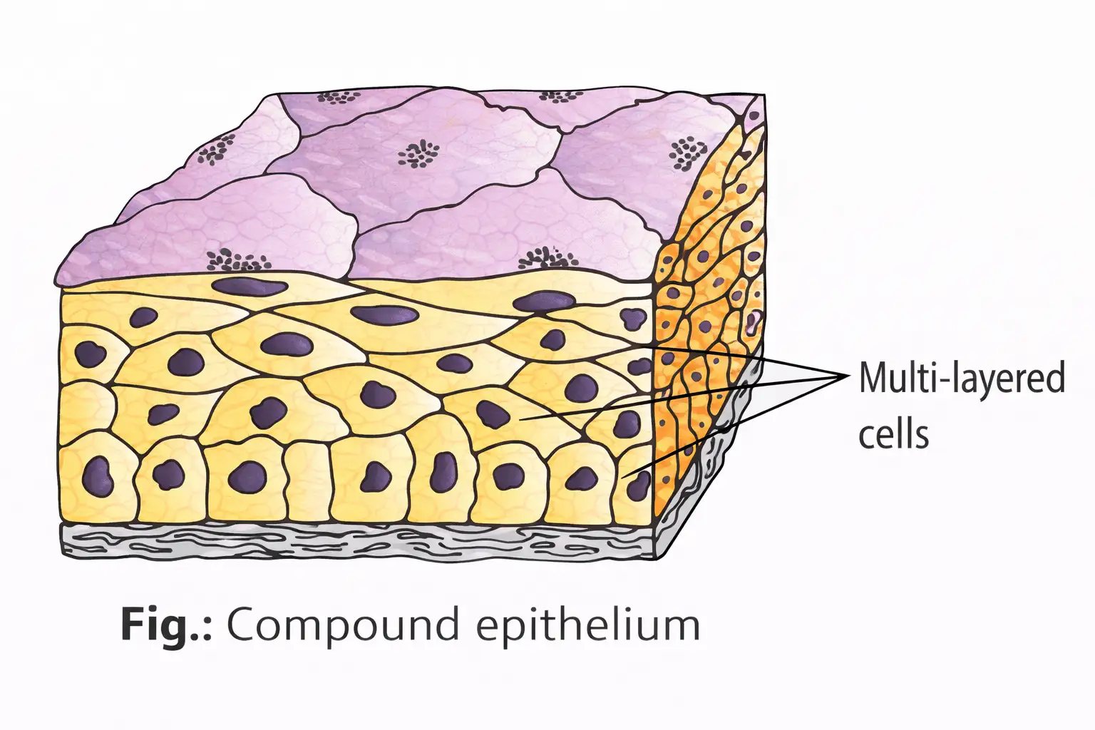
- Compound epithelium: More than one cell layer; mainly protects against mechanical and chemical stresses, with limited secretion and absorption.
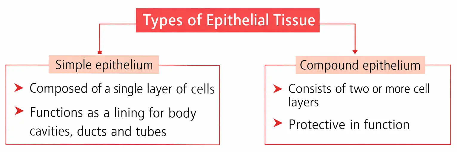
| Type | Structure and location | Main function / feature |
|---|---|---|
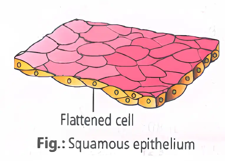
| Squamous (pavement) | Single thin layer of flattened cells with irregular boundaries; lines blood vessels and lung air sacs | Forms a diffusion boundary; also involved in protection, excretion, gas exchange and secretion of coelomic fluid |
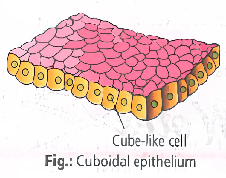
| Cuboidal | Single layer of cube-like cells; ducts of glands and kidney nephron tubules | Secretion and absorption; microvilli may increase absorptive surface area, e.g. PCT |
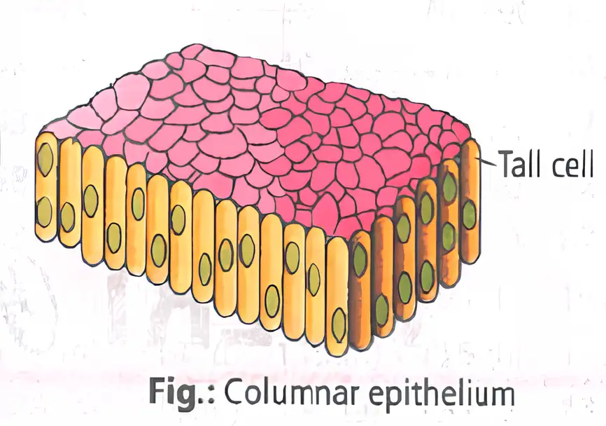
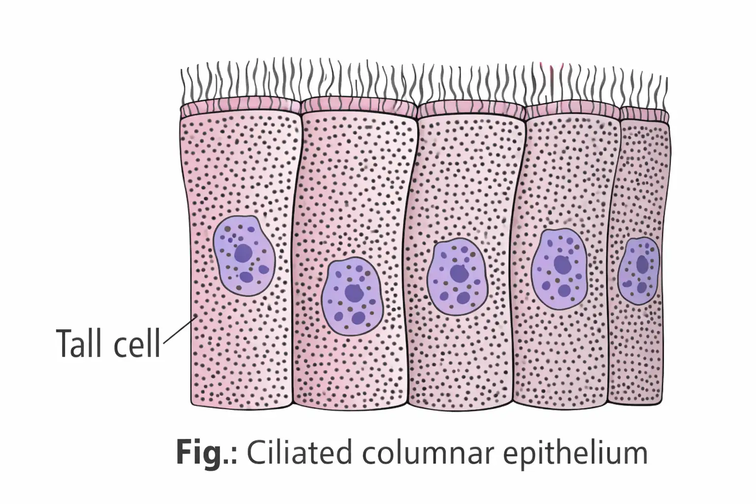
| Columnar | Single layer of tall, slender cells with nuclei near the base; lines stomach, intestine, gall bladder and bile duct | Intestinal microvilli form a brush border |
Ciliated epithelium
- Structure: Cuboidal or columnar epithelium with cilia on the free surface; it is a ciliated modification, not a fourth structural type of simple epithelium.
- Location and role: Lines hollow organs such as bronchioles and fallopian tubes; cilia move mucus, urine, eggs or cerebrospinal fluid in a particular direction.
Compound epithelium
- Locations: Dry skin surface; moist buccal cavity and pharynx; inner linings of salivary and pancreatic ducts.
- Main role: Protection from mechanical and chemical stress; secretion and absorption are limited.
Glandular epithelium and glands
Glandular epithelium — Epithelial cells specialised for secretion.
| Type | Organisation | Example |
|---|---|---|
| Unicellular | An isolated glandular cell | Goblet (mucous) cells in the alimentary canal |
| Multicellular | A cluster of glandular cells; may be tubular or saccular | Salivary glands |
| Type | Route | Examples |
|---|---|---|
| Exocrine | Secretion reaches an epithelial surface through ducts | Gastric, mucous, intestinal, salivary, sweat, sebaceous and mammary glands |
| Endocrine | Ductless; hormones are released directly into the fluid bathing the gland | Thyroid, adrenal and pituitary glands |
| Heterocrine | Has both exocrine and endocrine parts; secretion goes through a duct, while hormones enter blood and lymph | Pancreas and gonads |
| Mode | Example |
|---|---|
| Merocrine | Goblet cells |
| Apocrine | Mammary glands |
| Holocrine | Sebaceous glands |
Cell junctions
- Junctions: Special links in animal tissues provide structural and functional connections between cells.
| Junction | Function |
|---|---|
| Tight | Prevents substances from leaking across a tissue |
| Adhering | Cements neighbouring cells together |
| Gap | Connects cytoplasm of adjacent cells, enabling communication and rapid transfer of ions and molecules |
Connective tissue
Connective tissue — The most abundant and widely distributed tissue in complex animals; it links and supports other tissues and organs.
- Matrix: Cells secrete modified polysaccharides that accumulate between cells and fibres.
- Fibres: Except in blood, connective-tissue cells secrete collagen or elastin fibres, which provide strength, elasticity and flexibility.
- Range: Includes loose and dense connective tissues, cartilage, bone, adipose tissue and blood.
| Type | Arrangement |
|---|---|
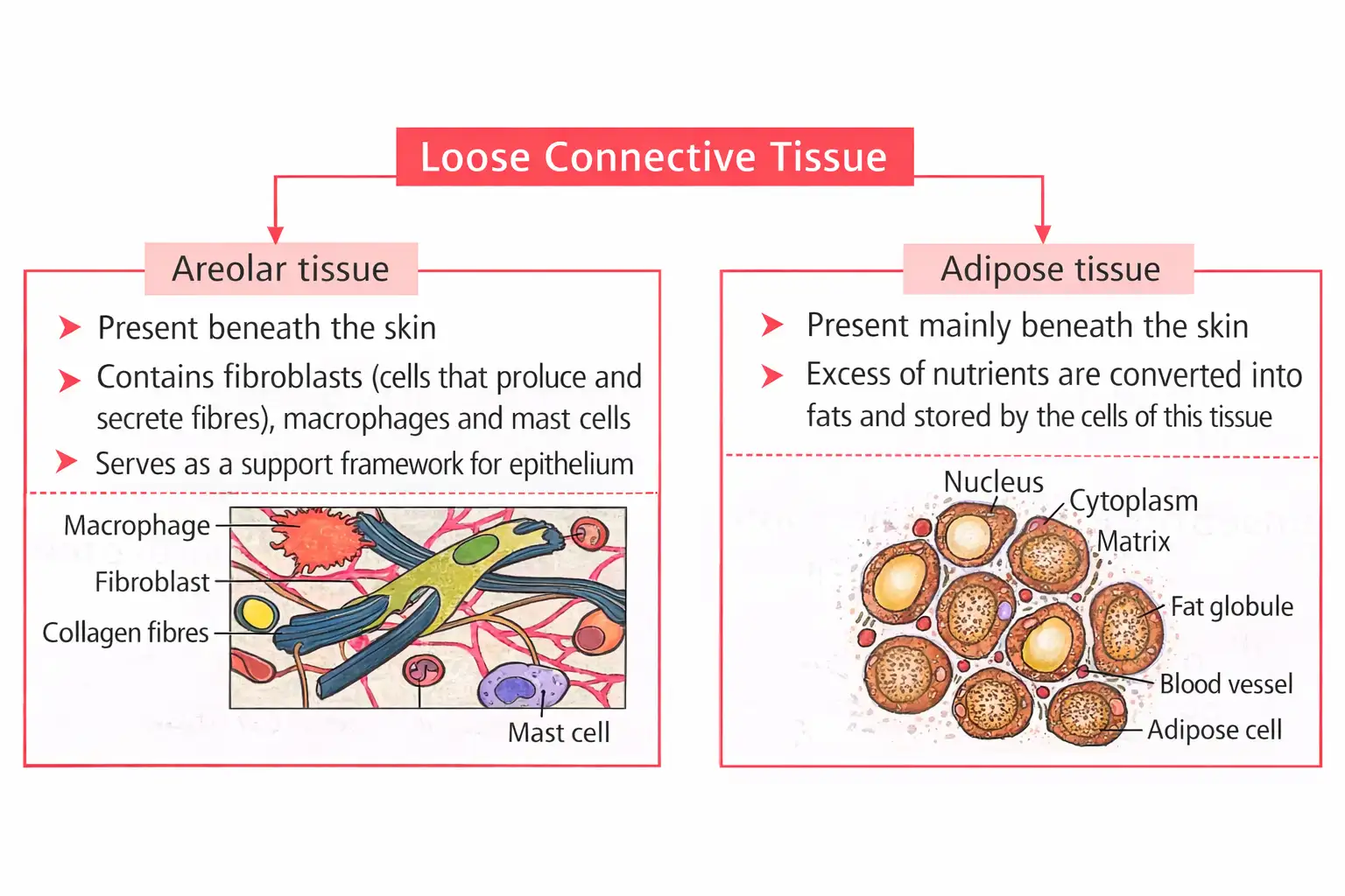
| Loose | Cells and fibres are loosely arranged in a semi-fluid ground substance; includes areolar and adipose tissue. |
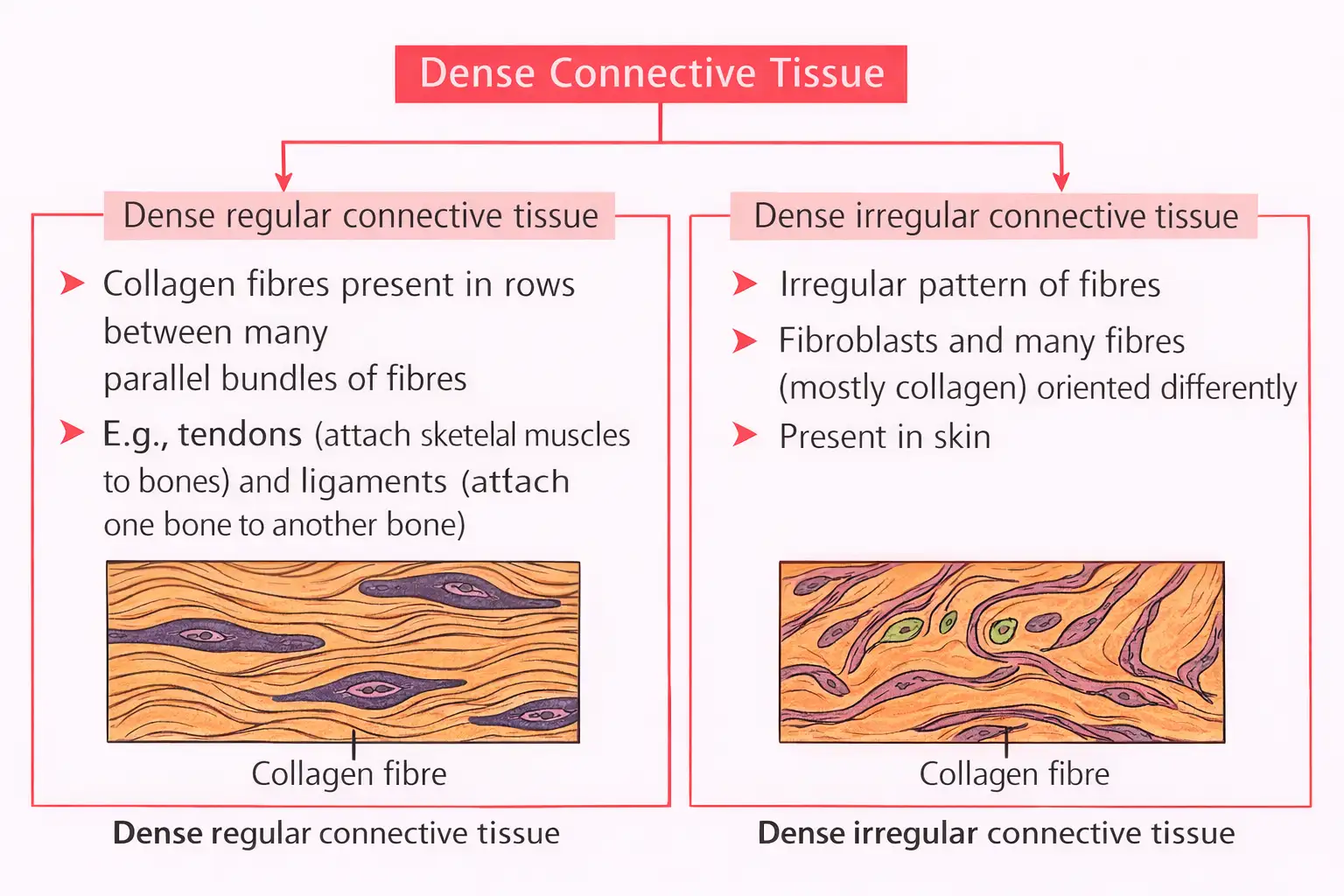
| Dense | Fibres and fibroblasts are compactly arranged; includes dense regular and dense irregular tissue. |
| Type | Structure and locations | Mechanical property |
|---|---|---|
| White fibrous | Mainly collagen fibres in bundles, with fibroblasts in rows; forms sheets in dermis, muscle and nerve sheaths, tunica adventitia of large vessels, periosteum, perichondrium, pericardium, dura mater and renal capsule | Great strength but limited flexibility |
| Yellow elastic | Thick, branched, loose network of yellow elastic fibres with irregularly scattered fibroblasts; forms ligaments that join bone to bone, and sheets in vessel walls, lungs, bronchioles, true vocal cords, laryngeal and tracheal cartilage | Considerable strength and remarkable elasticity |
Specialised connective tissues
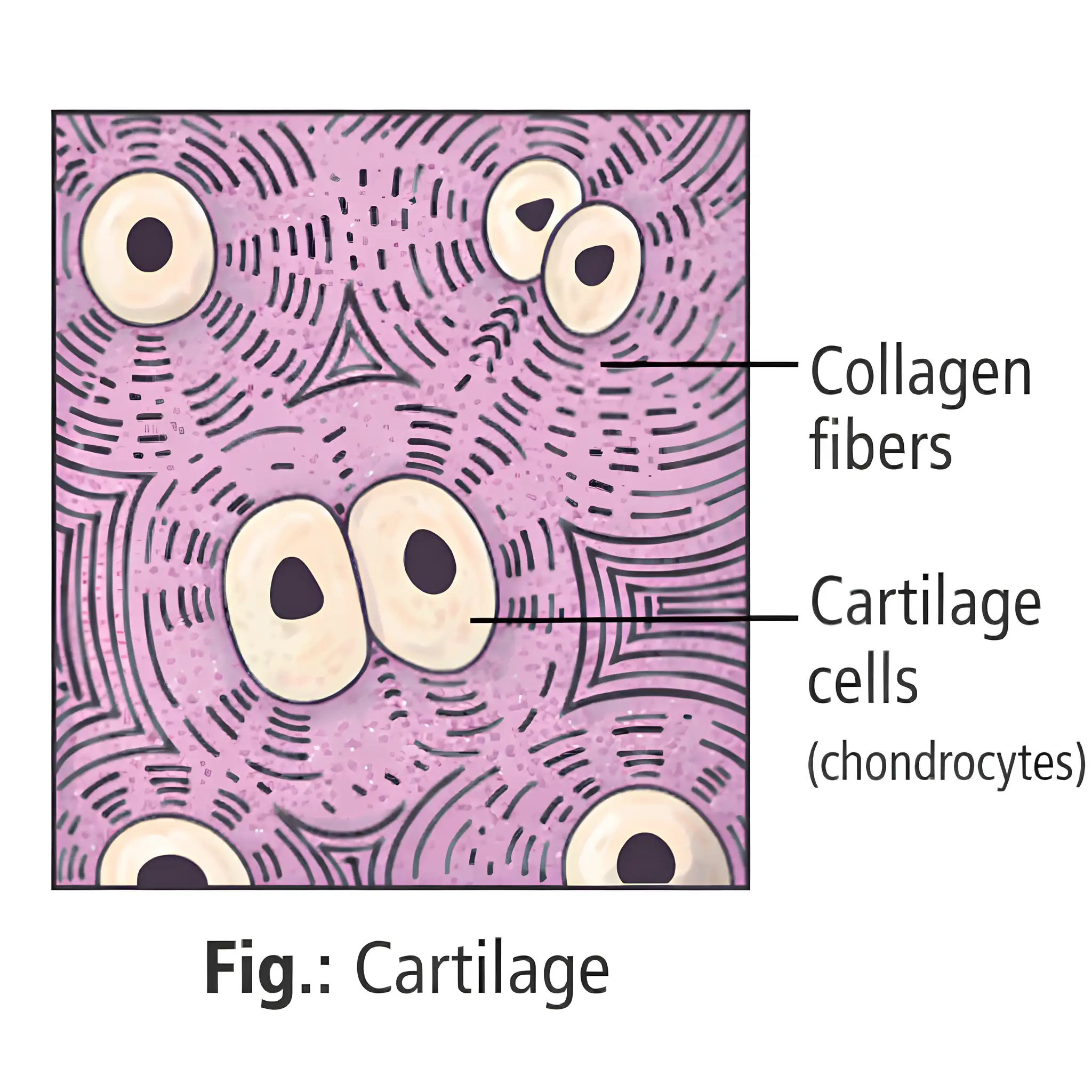
Cartilage — A soft skeletal tissue in which chondrocytes lie in cavities within their secreted matrix.
- Matrix: Solid but pliable; resists compression.
- Development: Most cartilage in vertebrate embryos is replaced by bone in adults.
| Type | Locations / distinguishing point |
|---|---|
| Hyaline | Most prevalent; articular surfaces of long bones, tracheal and bronchial rings, sternal parts of ribs, hyoid apparatus, nasal septum and part of larynx |
| White fibrous | Strongest cartilage; intervertebral discs and pubic symphysis |
| Yellow elastic fibrocartilage | Pinna and external auditory canal, Eustachian tubes, epiglottis and tip of nose |
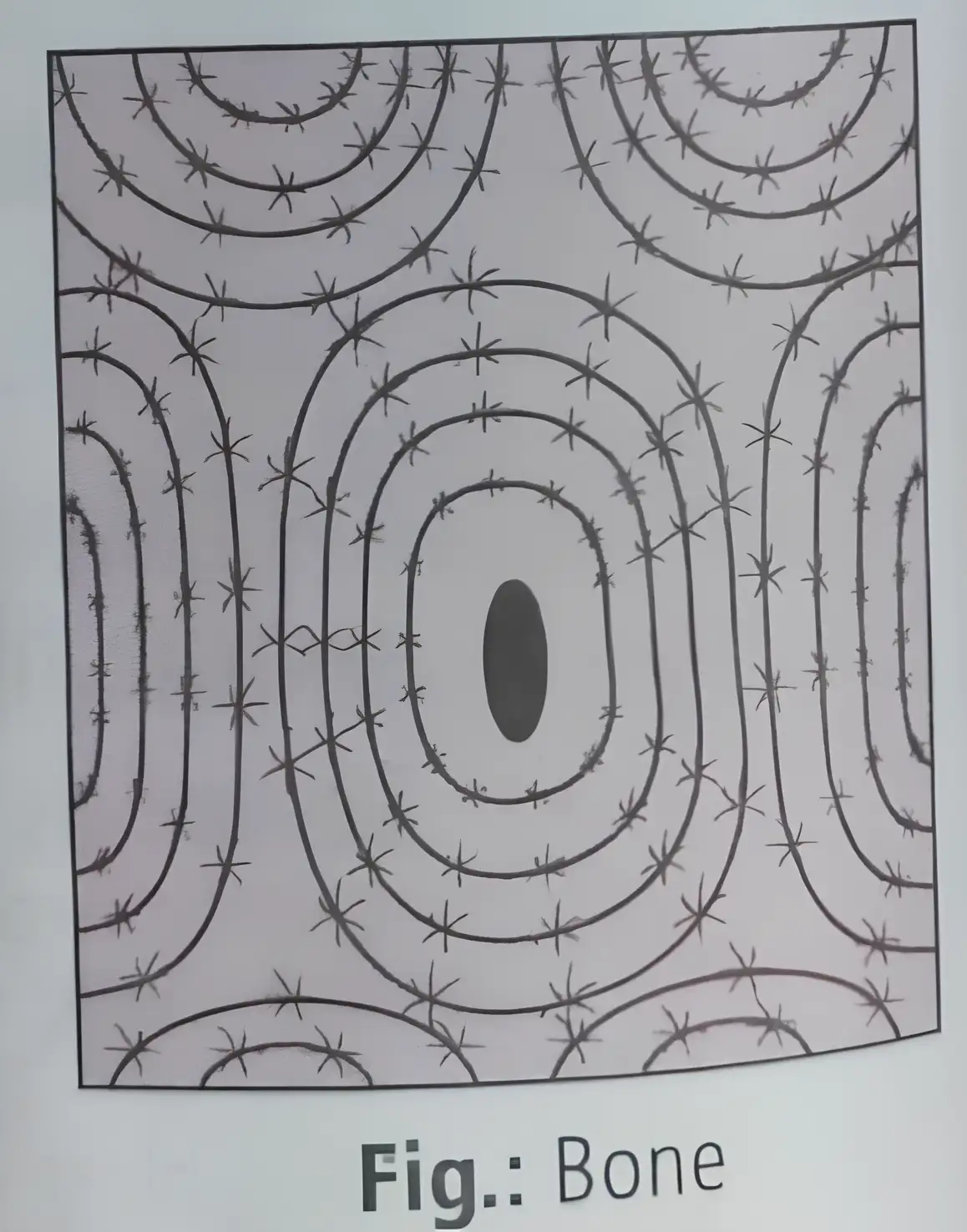
Bone — A hard skeletal connective tissue whose osteocytes occupy spaces called lacunae.
- Ground substance: Hard and non-pliable, rich in calcium salts and collagen fibres.
- Functions: Provides the body’s structural frame; supports and protects organs. Limb bones bear weight and work with skeletal muscles to produce movement.
- Red bone marrow: Site of blood-cell production.
- Osteon: A Haversian canal and its surrounding lamellae and osteocytes form a cylindrical unit called the Haversian system or osteon; absent from mammalian spongy bone.
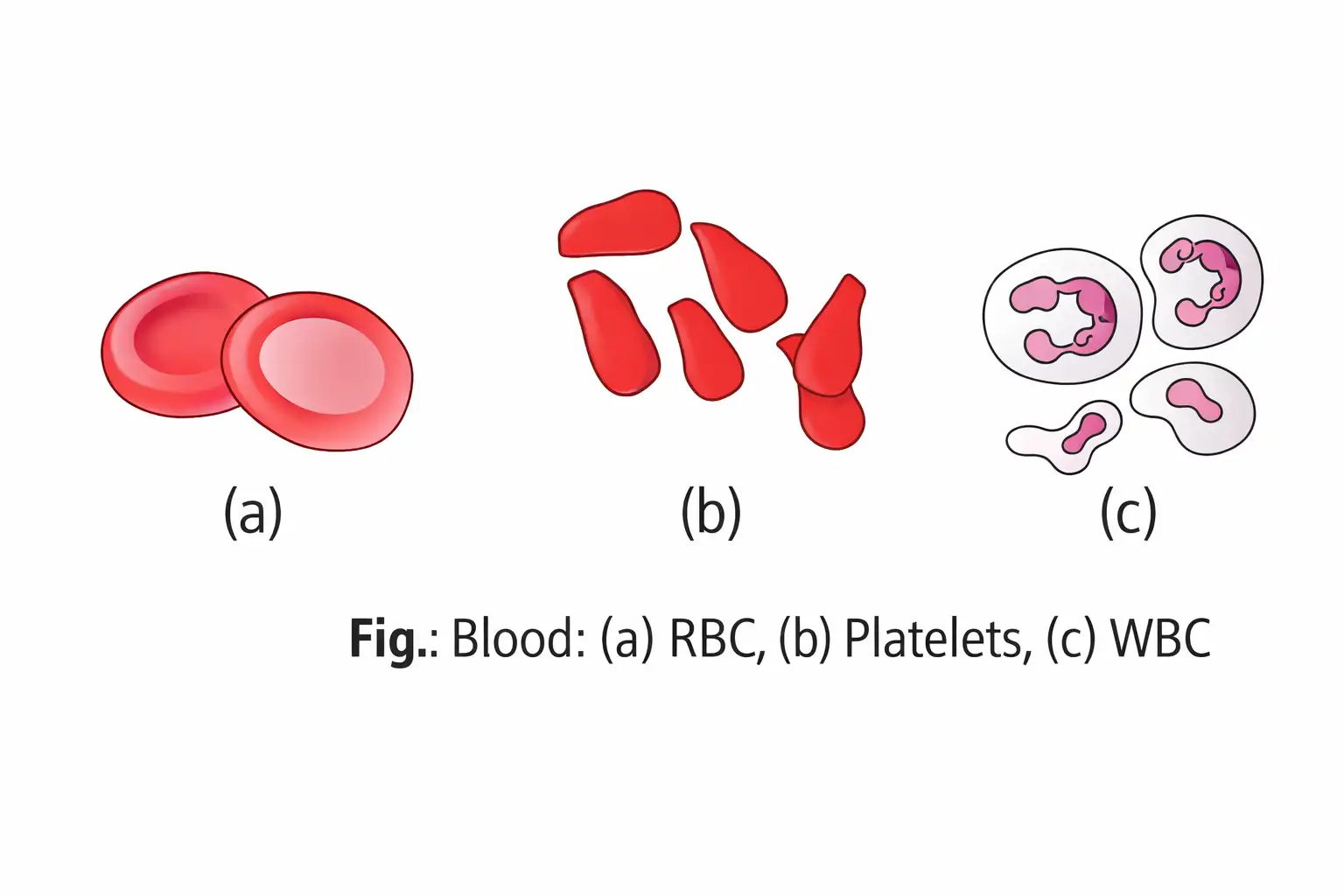
Blood — A fluid connective tissue made of plasma, red blood cells, white blood cells and platelets.
- Main role: Circulates through the body and transports substances.
Muscular tissue
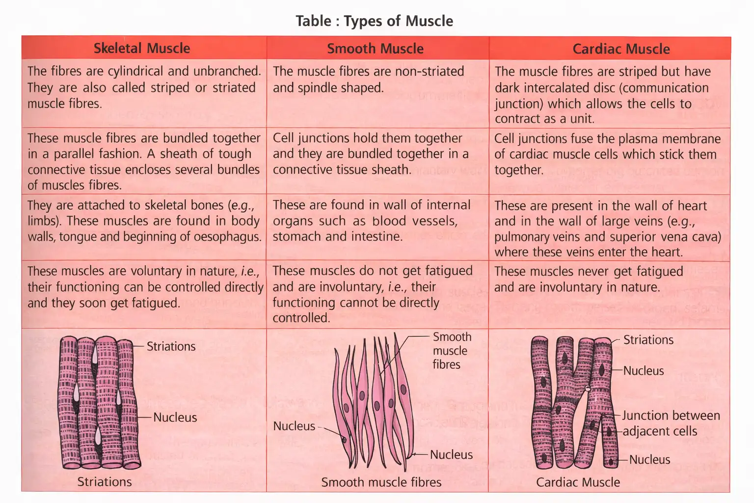
Muscular tissue — Tissue made of contractile muscle fibres that contract and relax to produce movement or maintain body posture.
- Structure: Muscle tissue consists of many long, cylindrical fibres; contractile filaments lie in parallel arrays and contain fine fibrils called myofibrils in the sarcoplasm.
- Action: Coordinated contraction and relaxation move body parts or maintain their position.
Neural tissue
Neuron — The main structural and functional unit of the neural system.
Neuron structure and neural tissue
- Cell body: Also called cyton or soma.
- Processes: Dendrites and an axon; the axon ends in terminal arborisations.
- Nerve fibre: An axon or dendrite covered by a sheath.
- Neuroglia: Make up more than 50% of neural-tissue volume and protect and support neurons.
- Response: Stimulation generates an electrical disturbance that travels rapidly along the neuron's plasma membrane.
| Type | Myelin sheath | Key feature |
|---|---|---|
| Medullated (myelinated) | Present, but interrupted at nodes of Ranvier | Nodes of Ranvier are the points where the sheath is absent |
| Non-medullated (non-myelinated) | Absent | Found in autonomic nerves |
Differentiate tight, adhering and gap junctions.
What is an osteon, and where is it absent?
Quick recap
- Animal tissues are epithelial, connective, muscular and neural, with characteristic origins and functions.
- A single cell performs basic life functions in unicellular organisms; in multicellular animals, specialised cells form tissues and organs.
- Simple epithelium has one layer and three structural types; cilia may occur on cuboidal or columnar cells.
- Glands are classified by cell number, route of secretion and mode of secretion; cell junctions prevent leakage, bind cells or enable communication.
- Connective tissue is the most abundant and widely distributed tissue; its types include loose, dense, cartilage, bone and blood.
- Cartilage resists compression; bone supports and protects, and red marrow produces blood cells.
- Cylindrical muscle fibres contract and relax; neurons conduct electrical disturbances while neuroglia support and protect them.
Organ and Organ System
Organisation of Tissues and Organs
Organ — A structure made of different tissues organised to perform specific functions.
- Example: The heart contains epithelial, connective, muscular and neural tissues.
- Organisation: Tissues form organs → organs form organ systems → organ systems make up an organism.
- Importance: This organisation enables efficient, coordinated activities among an organism’s cells.
| Term | Morphology | Anatomy |
|---|---|---|
| Study | Externally visible features; in animals, the external appearance of body parts and organs. | Internal structure of organs in animals. |
Cockroach: Identity and General Features
- Scientific name: *Periplaneta americana*; Class Insecta, Phylum Arthropoda.
- Habit: Brown or black-bodied, nocturnal and omnivorous.
Cockroach Morphology
- Body regions: The segmented body is divided into head, thorax and abdomen.
- Exoskeleton: The hard chitinous covering has plates called sclerites—dorsal tergites and ventral sternites—joined by flexible arthrodial membrane.
- Head: Triangular and formed by fusion of six segments; the flexible neck allows movement. The head bears compound eyes and sensory-receptor-rich antennae.
- Thorax: Prothorax, mesothorax and metathorax each bear one pair of walking legs; each leg consists of segments called podomeres.
- Wings: Two pairs arise from the mesothorax and metathorax. Dark, opaque, leathery forewings cover the transparent, membranous hindwings at rest; hindwings are used for flight.
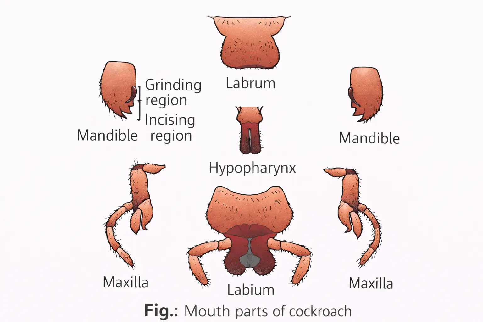
Mouthparts
- Type: Mouthparts are adapted for chewing and biting.
- Parts: Labrum (upper lip), labium (lower lip), one pair each of mandibles and maxillae, and the hypopharynx, which acts like a tongue.
Cockroach Anatomy: Abdomen and External Sex Differences
- Abdomen: Both sexes have 10 segments.
- Female pouch: The boat-shaped seventh sternum, with the eighth and ninth sterna, forms the brood or genital pouch. Its anterior part contains the female gonopore, spermathecal pores and collaterial glands.
- Male pouch: The hind-end genital chamber is bounded dorsally by the ninth and tenth terga and ventrally by the ninth sternum; it contains the anus, male genital pore and gonapophyses.
- Anal structures: Males have a pair of short, thread-like anal styles, absent in females. Both sexes have a pair of jointed anal cerci on the tenth segment.
Digestive System
Alimentary canal — A tube divided into three regions: foregut, midgut and hindgut.
| Region | Parts and functions | Key features |
|---|---|---|
| Foregut | Mouth → pharynx → narrow oesophagus → crop (food storage) → gizzard or proventriculus (grinding). | Gizzard has six highly chitinous teeth/plates. |
| Midgut | Short, narrow, coiled tube; digestion is aided by hepatic caecae. | Six to eight blind hepatic caecae at the foregut–midgut junction secrete digestive juice. |
| Hindgut | Ileum → colon → rectum; rectum opens through the anus. | Has six rectal glands and is more permeable to water than the foregut. |
Digestive-System Junctions and Lining
- Excretion at junction: About 100–150 thin Malpighian tubules at the midgut–hindgut junction remove excretory products from haemolymph.
- Lining: Foregut and hindgut are ectodermal and lined with cuticle; the endodermal midgut lacks cuticle.
Blood Vascular System
- Type: Open circulatory system; vessels open into the haemocoel, where haemolymph bathes visceral organs.
- Haemolymph: Colourless plasma and haemocytes; it lacks respiratory pigment and transports food and metabolic wastes, not respiratory gases.
- Heart: An elongated muscular tube divided into funnel-shaped chambers, with ostia on either side.
- Flow: Haemolymph enters the heart from sinuses through ostia and is pumped anteriorly back into the sinuses.
- Control: The cockroach heart is neurogenic.
Respiratory System
- Air entry: A tracheal network opens outside through 10 pairs of lateral spiracles, guarded by bristles or hairs that keep out dirt.
- Gas transport: Tracheae branch into tracheoles, which carry oxygen to body parts; gas exchange occurs by diffusion.
- Carbon dioxide: Only a small amount leaves through spiracles; most exits through the body’s cuticular covering.
Excretory System
- Main organs: Glandular and ciliated Malpighian tubules absorb nitrogenous wastes and convert them into uric acid.
- Water and salts: Malpighian tubules also reabsorb certain salts and water.
- Other structures: Fat body, nephrocytes and uricose glands also assist in excretion.
- Excretory type: Cockroaches are uricotelic because they excrete nitrogenous waste as uric acid.
Nervous System and Sense Organs
- Divisions: Central, peripheral and sympathetic nervous systems.
- Ventral nerve cord: Segmentally arranged fused ganglia are joined by paired longitudinal connectives; there are nine ganglia—three thoracic and six abdominal.
- Brain: The bilobed supra-oesophageal ganglion supplies nerves to the antennae and compound eyes.
- Compound eyes: Each dorsal eye has about 2,000 hexagonal ommatidia, which together produce mosaic vision.
- Vision: Mosaic vision suits nocturnal activity; it has greater sensitivity but lower resolution.
- Other sense organs: Antennae, maxillary palps, labial palps and anal cerci.
Reproductive System
- Sexes: Cockroaches are dioecious; males and females are separate individuals.
| Point | Male | Female |
|---|---|---|
| Gonads | A pair of three-lobed testes in abdominal segments 4–6; each has numerous whitish, transparent follicles. | Ovaries in abdominal segments 2–6; each contains ovarian tubules (ovarioles) with a chain of developing ova. |
| Ducts and storage | Vasa deferentia lead from the testes and connect to the ejaculatory duct through seminal vesicles. The ejaculatory duct opens into the male gonopore. Sperm are stored in the seminal vesicles as bundles called spermatophores. | Oviducts unite into a median oviduct that opens into the genital chamber; a pair of spermathecae in segment 6 also opens there. |
| Accessory structures | A mushroom-shaped accessory gland lies in abdominal segments 6–7; external genitalia are gonapophyses (phallomeres). | Paired collaterial glands lie behind the ovaries; their secretion forms the egg case (ootheca). |
Fertilisation and Development
- Sperm transfer: Sperms are transferred in spermatophores during copulation.
- Ootheca: Fertilised eggs are enclosed in reddish-brown capsules, which are dropped or glued to a humid surface.
- Egg number: A female produces about 9–10 oothecae on average, each containing 14–16 eggs.
- Development: Paurometabolous development includes a nymphal stage; the nymph moults about 13 times before reaching adulthood.
Economic and Health Importance
- Pest species: Some cockroaches damage food and contaminate it with foul-smelling excreta.
- Disease transmission: Cockroaches can spread various bacterial diseases by contaminating food.
How does the cockroach alimentary canal differ among its three regions?
Why is the cockroach described as uricotelic, and what is its main excretory organ?
Quick recap
- Tissues form organs, organs form organ systems, and organ systems form an organism.
- Cockroaches have a chitinous exoskeleton, segmented body and chewing-biting mouthparts.
- The open circulatory system uses haemolymph, which does not transport respiratory gases.
- Tracheae and tracheoles deliver oxygen; Malpighian tubules excrete uric acid.
- Nine ventral ganglia support the nervous system; compound eyes provide sensitive, low-resolution mosaic vision.
- Cockroaches are dioecious; eggs develop in oothecae, and development proceeds through a moulting nymph.
Frog
General Features
Morphology
Anatomy: Digestive System
Anatomy: Respiratory System
Anatomy: Circulatory System
Anatomy: Excretory System
Control and Coordination
Endocrine System
Nervous System and Sense Organs
Reproductive System
Economic Importance
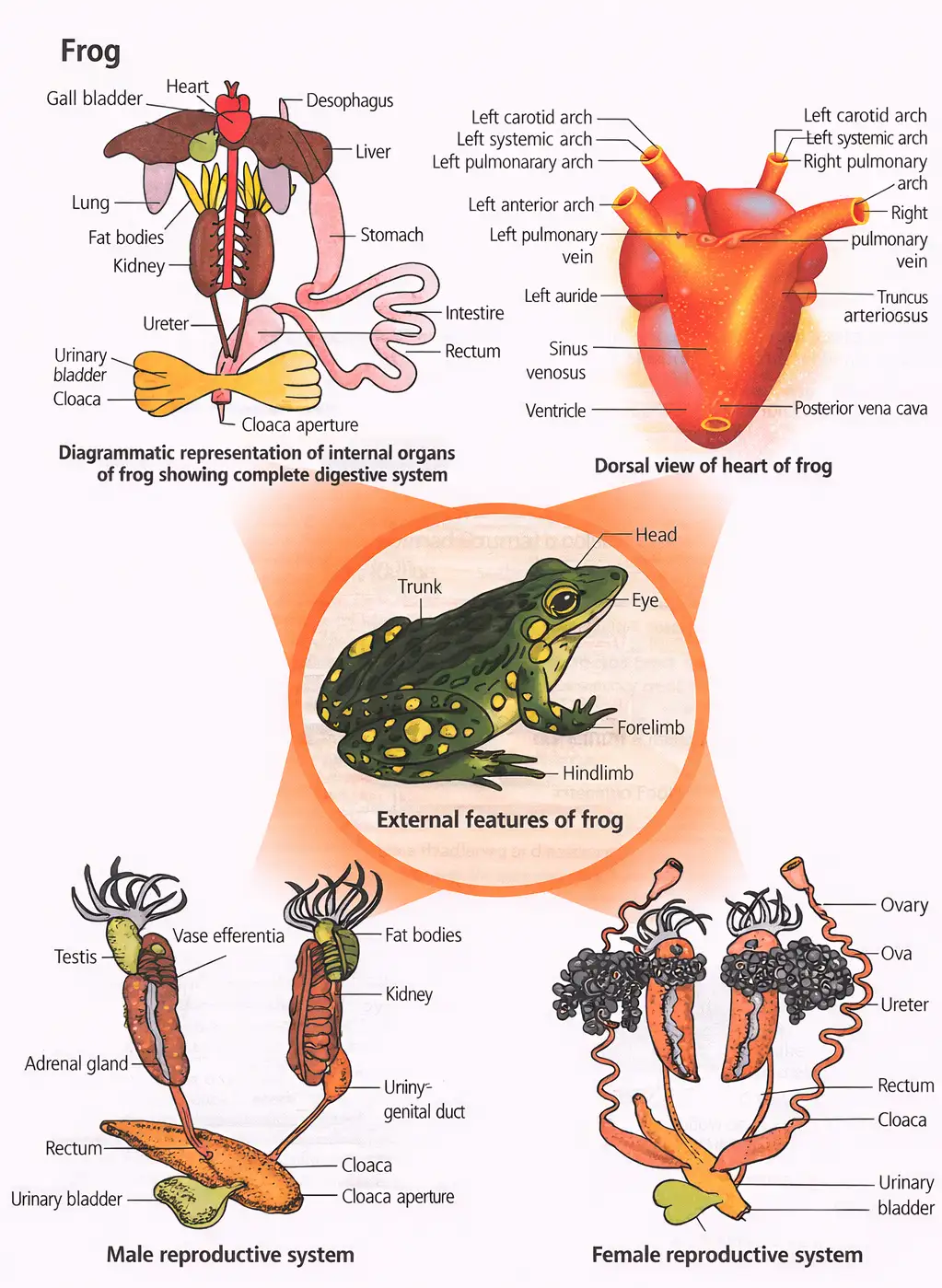
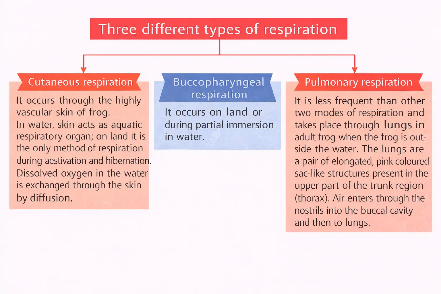
Describe the respiratory system of an adult frog as stated in this lecture.
How is the frog’s heart organised?
Summarise fertilisation and development in frogs.
Quick recap
- Frogs are amphibian chordates, poikilothermic and capable of aestivation and hibernation.
- Their moist skin absorbs water; males and females differ in vocal sacs, copulatory pads, body build and colour.
- The short digestive canal leads to the cloaca; the intestine completes digestion and absorbs nutrients through villi and microvilli.
- Frog circulation is closed, with a myogenic, three-chambered heart and hepatic and renal portal systems.
- Frogs are ureotelic; male ureters act as urinogenital ducts, while female oviducts open separately into the cloaca.
- Fertilisation is external in water; the tadpole undergoes metamorphosis into an adult.
NEET Zoology — ALL CHAPTERS
1
Structural Organisation in Animals
2
Breathing and Exchange of Gases
3
Body Fluids and Circulation
4
Excretory Products and their Elimination
5
Locomotion and Movement
6
Neural Control and Coordination
7
Human Reproduction
8
Reproductive Health
9
Evolution
10
Microbes in Human Welfare
11
Organism and Population
12
Ecosystem
13
Biodiversity and Conservation