NEET Ug . NEET Zoology . Zoology . Human Reproduction
Chapter 7: Human Reproduction
Ch 7
NEET Zoology
NEET Ug
The Male Reproductive System
Human Reproductive Events and Organization
Overview
- Human reproduction is sexual; humans are viviparous, so the developing young is born from the mother.
- Gametogenesis is the formation of gametes; insemination is the transfer of sperm into the female genital tract.
- Fertilisation is the fusion of male and female gametes, forming a zygote; the zygote develops into a blastocyst, which attaches to the uterine wall during implantation.
- Gestation is embryonic development; parturition is delivery of the baby. Sequence: gametogenesis → insemination → fertilisation → zygote → blastocyst formation and implantation → gestation → parturition.
- Reproductive events begin after puberty. Primary sex organs produce gametes and sex hormones.
- Secondary sex organs neither produce gametes nor secrete sex hormones; they perform supporting functions, including gamete conduction.
- Secondary sexual characters are external features that distinguish the sexes.
Male reproductive system — A pelvic system made up of a pair of testes, accessory ducts, accessory glands and external genitalia.
Main components
- Testes lie outside the abdominal cavity in a pouch called the scrotum.
- Testes are the primary sex organs: they produce sperm and secrete male sex hormones.
Scrotum and Temperature Regulation
Structure and role
- Scrotum connects with the abdomen or pelvis through the inguinal canals.
- Spermatic cord passes through the inguinal canal and contains the spermatic artery, vein and nerve bound together by connective tissue.
- Temperature: the scrotum keeps the testes near \(34^\circ\text{C}\)—about \(2\)–\(2.5^\circ\text{C}\) below the internal body temperature of \(37^\circ\text{C}\)—for optimal sperm production.
- Temperature control: scrotal receptors respond to temperature; testes move towards or away from the pelvis to conserve or disperse heat.
- Cold conditions: testes move closer to the pelvis to absorb body heat. Excess heat: they move away, increasing exposed surface area for heat loss.
- Cremaster and dartos muscles contract or relax to adjust the position of the testes.
Testis and Seminiferous Tubules
Testis structure
- Adult testis: oval, about \(4\)–\(5\text{ cm}\) long and \(2\)–\(3\text{ cm}\) wide; covered by tunica vaginalis, tunica albuginea and tunica vasculosa.
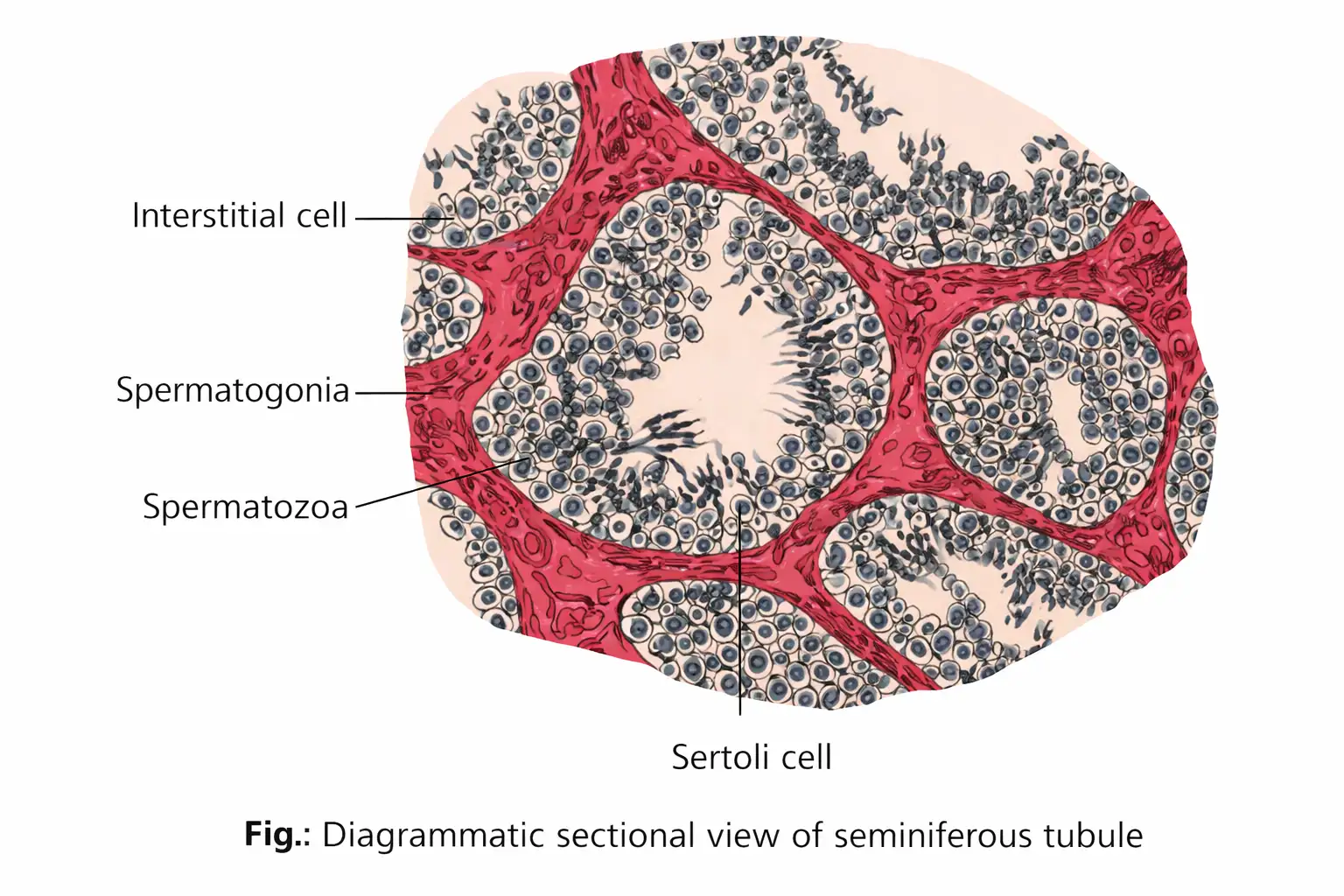
- Testicular lobules: each testis has about \(250\) compartments; each contains \(1\)–\(3\) highly coiled seminiferous tubules.
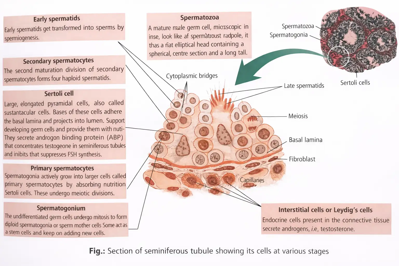
- Seminiferous tubules are the site of spermatogenesis and are lined by male germ cells (spermatogonia) and Sertoli cells.
- Male germ cells develop into sperm through meiotic divisions; Sertoli cells provide nutrition to the developing germ cells.
- Interstitial spaces between tubules contain small blood vessels, Leydig cells and other immune cells, including lymphocytes, macrophages and mast cells.
- Leydig cells synthesise and secrete androgens, the testicular hormones.
- Two testis functions: sperm production and secretion of male sex hormones.
Male Accessory Ducts
Sperm transport pathway
- Seminiferous tubules → rete testis
- Rete testis → vasa efferentia
- Vasa efferentia → epididymis
- Epididymis → vas deferens, which ascends into the abdomen and loops over the urinary bladder
- Vas deferens joins the duct from the seminal vesicle to form the ejaculatory duct → urethra → external urethral meatus
Duct functions
- Accessory ducts store and transport sperm from the testes to the outside through the urethra.
- Epididymis lies along the posterior surface of each testis and has three regions: caput, corpus and cauda.
- Epididymis stores mature sperm until ejaculation.
- Urethra begins at the urinary bladder, passes through the penis and opens externally at the urethral meatus.
External Genitalia: Penis
Structure and functions
- Penis is the male copulatory organ and serves as both a reproductive organ and a urinary duct.
- Erectile tissue: three cylindrical masses—two dorsal corpora cavernosa and one ventral corpus spongiosum—are surrounded by fibrous tissue.
- Corpus spongiosum surrounds the penile urethra and expands at the tip to form the glans penis.
- Prepuce (foreskin) is a loose fold of skin covering the glans penis.
Male Accessory Glands
| Gland | Location / structure | Secretion and composition | Main functions |
|---|---|---|---|
| Seminal vesicles (paired) | Sac-like structures in the pelvis near the base of the bladder; ducts join the vasa deferentia. | Alkaline secretion; about \(60\)–\(70\%\) of semen. Contains fructose, prostaglandins, clotting proteins and calcium. | Fructose supplies sperm energy; prostaglandins stimulate uterine contractions that aid sperm movement towards the oviduct; clotting proteins promote semen coagulation after ejaculation. |
| Prostate gland | Large, chestnut-shaped gland surrounding the urethra. | Slightly alkaline, milky fluid; about \(20\)–\(30\%\) of semen. Contains citric acid, enzymes such as acid phosphatase and amylase, and prostaglandins. | Citric acid nourishes sperm; prostaglandins activate sperm movement. The fluid nourishes and activates spermatozoa. |
| Bulbourethral glands (Cowper's glands; paired) | Pea-sized glands on either side of the membranous urethra; ducts open into the urethra. | Alkaline fluid and mucus. | Alkaline fluid neutralises urine acidity in the urethra; mucus lubricates the end of the penis and the urethral lining. |
Semen
Semen — A mixture of sperm from the testes and secretions from the seminal vesicles, prostate gland and Cowper's glands.
Key properties
- Ejaculation: semen is expelled from the penis; a single ejaculation may contain \(200\)–\(300\) million sperm.
- pH: semen is alkaline, with a pH of \(7.35\) to \(7.50\).
- Functions: provides a fluid medium for sperm transport, nourishes sperm and helps neutralise acidity in the male urethra, protecting sperm.
Related Conditions
| Condition | Definition / cause |
|---|---|
| Cryptorchidism | Failure of one or both testes to descend into the scrotum; associated with deficient testosterone secretion by the fetal testes. |
| Inguinal hernia | Tearing of inguinal tissue can allow part of the intestine to protrude into the scrotum. |
Trace the pathway followed by sperm from the seminiferous tubules to the exterior.
State the two functions of the testes and identify the cells responsible for them.
Quick recap
- Gametogenesis forms gametes; insemination transfers sperm; fertilisation forms a zygote; implantation, gestation and parturition follow.
- Primary sex organs produce gametes and hormones; secondary sex organs do neither, but support functions such as gamete conduction.
- The testes produce sperm and androgens; Sertoli cells nourish developing germ cells, while Leydig cells secrete androgens.
- The scrotum maintains testes near \(34^\circ\text{C}\), below normal internal body temperature.
- Sperm travel through the rete testis, vasa efferentia, epididymis, vas deferens, ejaculatory duct and urethra.
- Seminal vesicles, prostate and bulbourethral glands contribute secretions to alkaline semen, which transports and nourishes sperm.
The Female Reproductive System
Overall Organisation
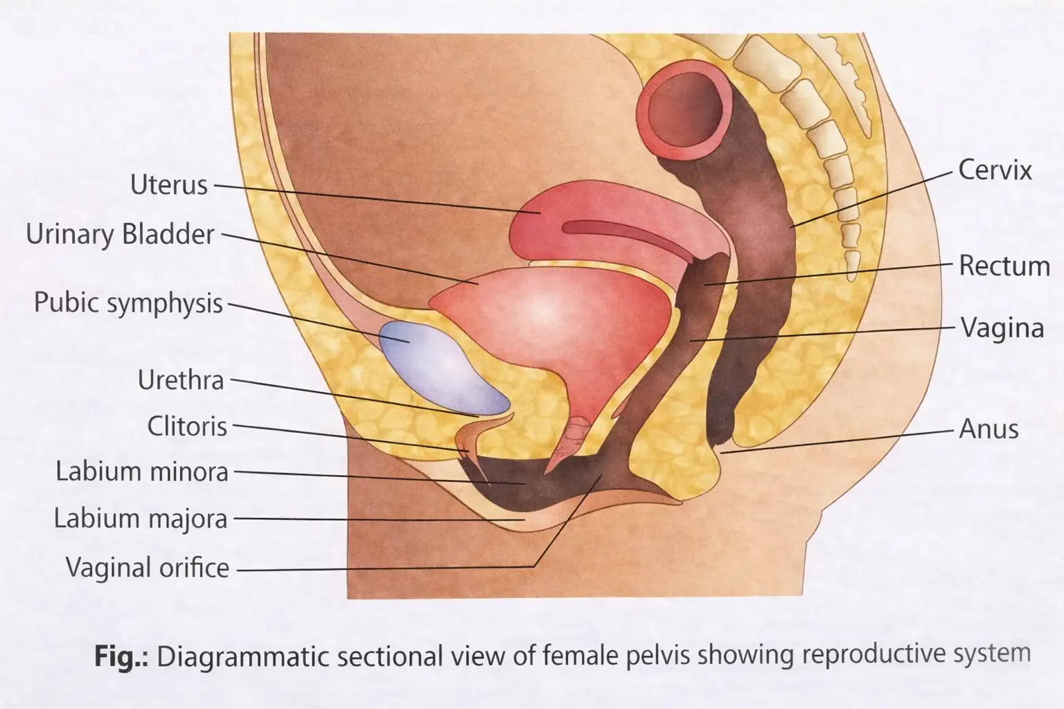
Female reproductive system — The female reproductive system comprises the ovaries, accessory ducts, external genitalia and mammary glands, which work together in reproduction and child care.
Ovaries
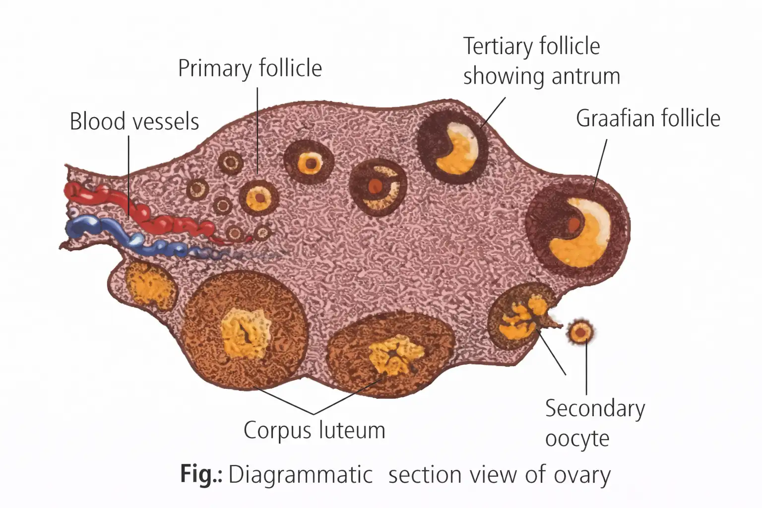
Ovaries — A pair of almond-shaped female gonads that produce ova through oogenesis.
Female Accessory Ducts
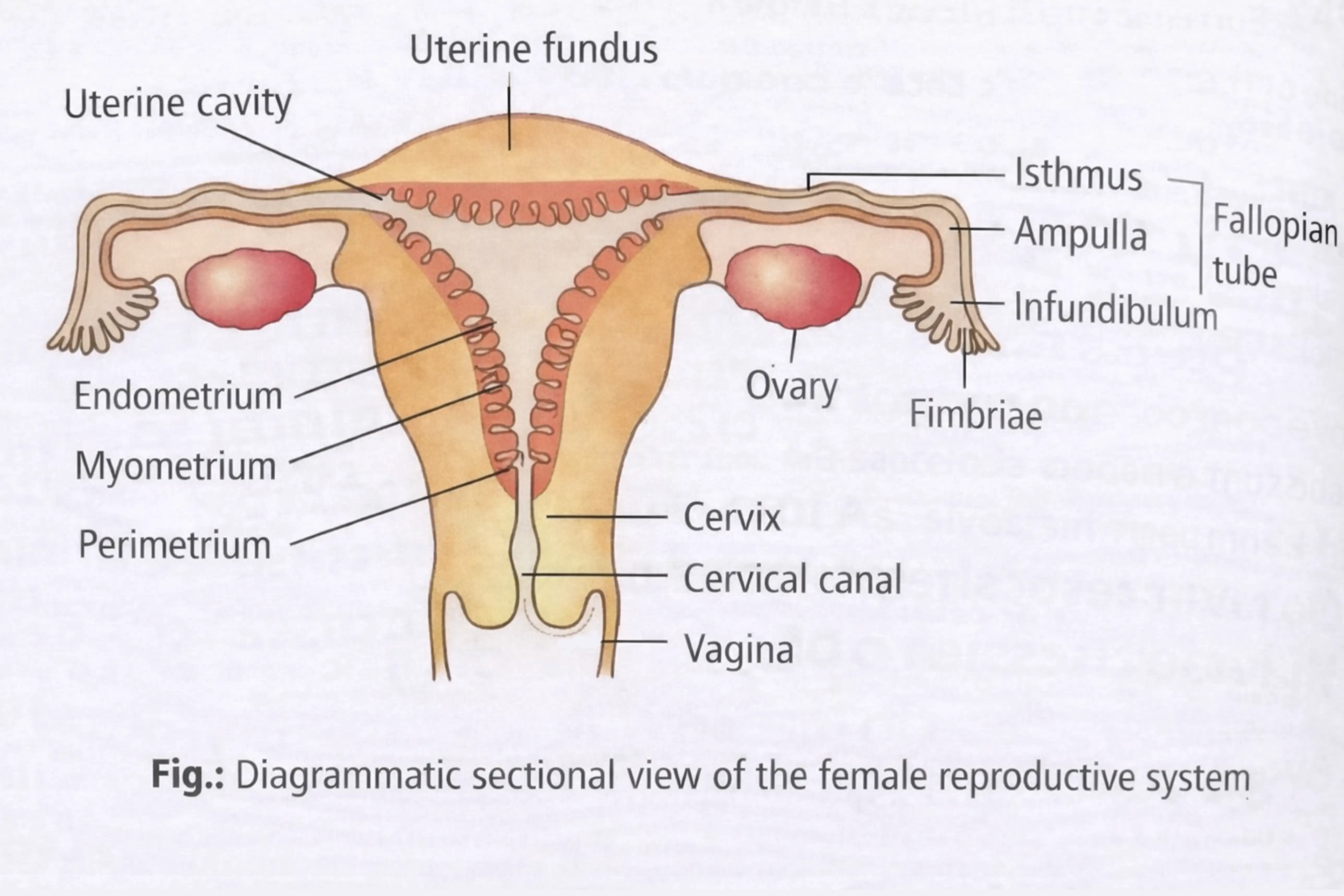
Female accessory ducts — The oviducts, uterus and vagina form the female accessory ducts.
Uterus — A single, inverted pear-shaped organ, also called the womb, supported by ligaments attached to the pelvic wall.
| Layer | Position and structure | Key feature |
|---|---|---|
| Perimetrium | Thin outer membrane | External layer |
| Myometrium | Thick middle smooth-muscle layer | Contracts strongly during delivery |
| Endometrium | Inner glandular lining of the uterine cavity | Undergoes cyclical changes during the menstrual cycle |
Vagina — A tube about 10 cm long that extends from the cervix to the exterior of the body.
External Genitalia
Female external genitalia — The external genitalia include the mons pubis, labia majora, labia minora, hymen and clitoris.
Female Accessory Glands
Vestibular glands — Vestibular glands are mucus- or fluid-secreting glands around the urethral and vaginal openings.
| Type and other name | Location | Secretion and relation |
|---|---|---|
| Lesser vestibular glands; paraurethral glands; glands of Skene | Numerous small glands on both sides of the urethral opening | Secrete mucus; homologous to the male prostate |
| Greater vestibular glands; Bartholin's glands | A pair, one on each side of the vaginal opening | Secrete viscid lubricating fluid during sexual arousal; homologous to the male Cowper's glands |
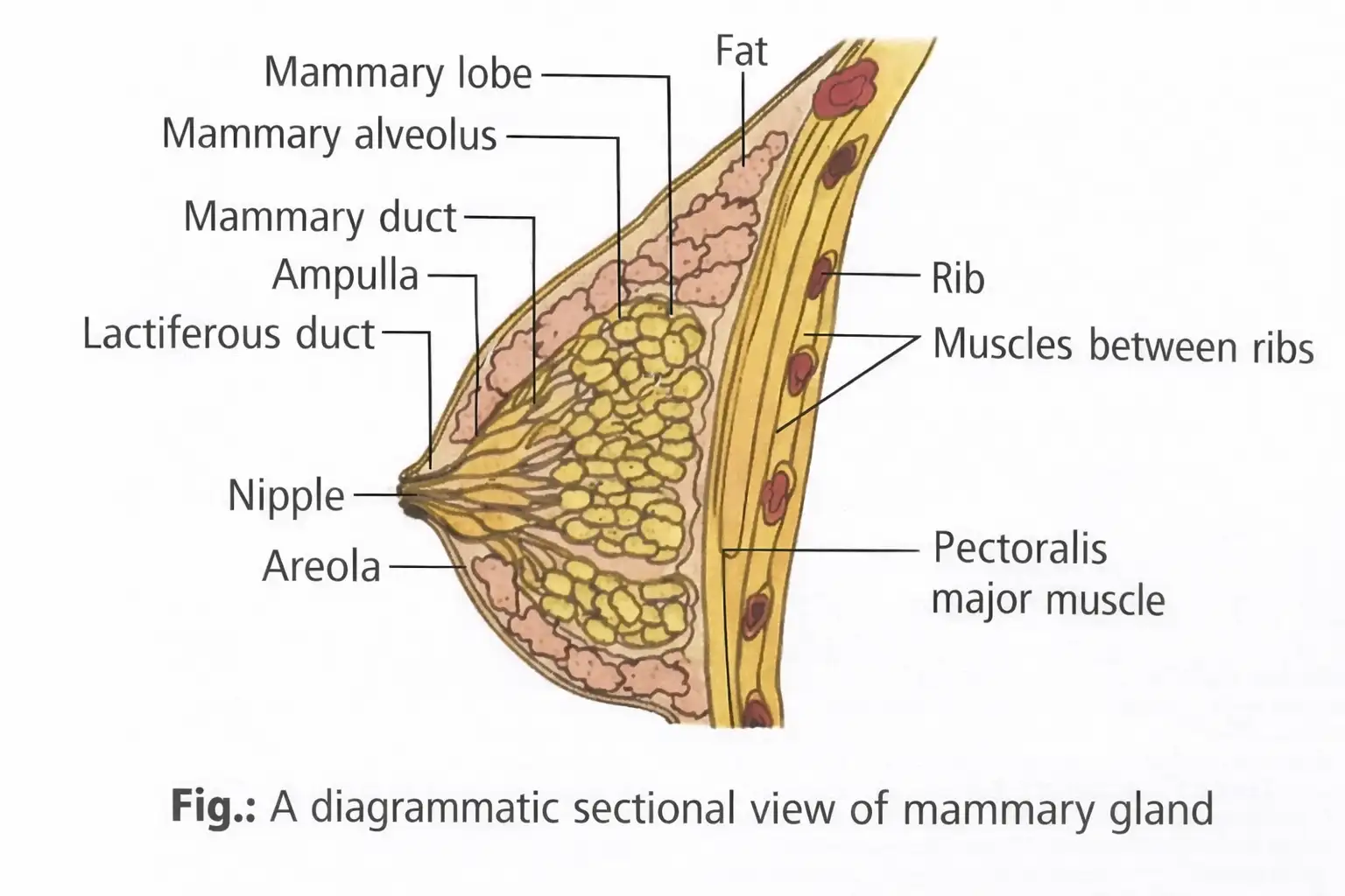
Mammary glands — Paired breast glands containing glandular tissue and variable amounts of fat; functional mammary glands produce and deliver milk.
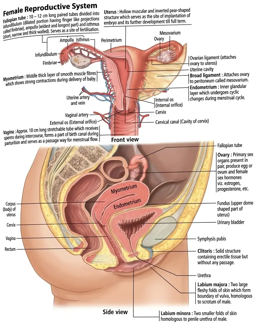
Name the three parts of the oviduct in order from the ovary towards the uterus.
Which uterine layer contracts strongly during delivery, and which changes cyclically during the menstrual cycle?
Quick recap
- The reproductive system includes paired ovaries and oviducts, uterus, cervix, vagina, external genitalia and mammary glands.
- Ovarian stroma has a dense cortex and a vascular central medulla.
- The oviduct runs from infundibulum to ampulla to isthmus; fimbriae help collect the ovum.
- The uterus has perimetrium, myometrium and endometrium; the cervical canal and vagina form the birth canal.
- The hymen is not a reliable indicator of sexual experience.
- Skene's glands secrete mucus; Bartholin's glands secrete lubricating fluid.
- Milk pathway: alveoli → mammary tubules → mammary duct → mammary ampulla → lactiferous duct.
Gametogenesis
Gametogenesis — The formation of male and female gametes—sperms and ova—from germ cells in the testes and ovaries.
Spermatogenesis
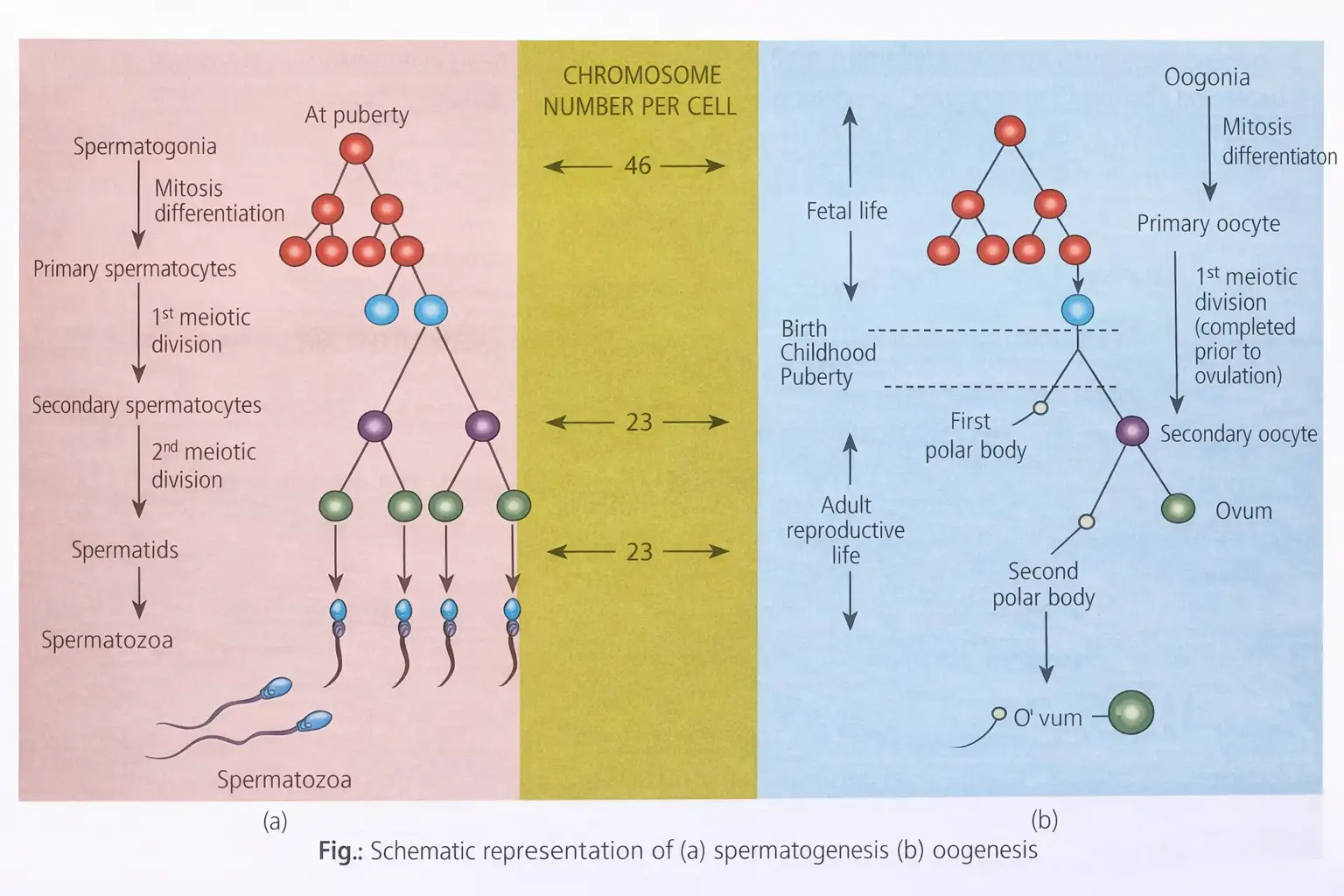
Spermatogenesis — The formation of sperm from spermatogonia in the seminiferous tubules of the testes; it begins at puberty.
| Phase | Key event |
|---|---|
| Multiplication | Diploid spermatogonia divide by mitosis, producing many spermatogonia. |
| Growth | Each diploid spermatogonium grows into a primary spermatocyte, nourished by supporting cells. |
| Maturation | Meiosis I forms two haploid secondary spermatocytes; meiosis II forms spermatids. |
Chromosome outcome
- Primary spermatocyte: Diploid, with \(46\) chromosomes in humans.
- Products: One primary spermatocyte produces four haploid spermatids, each with \(23\) chromosomes.
Sperm Formation and Structure
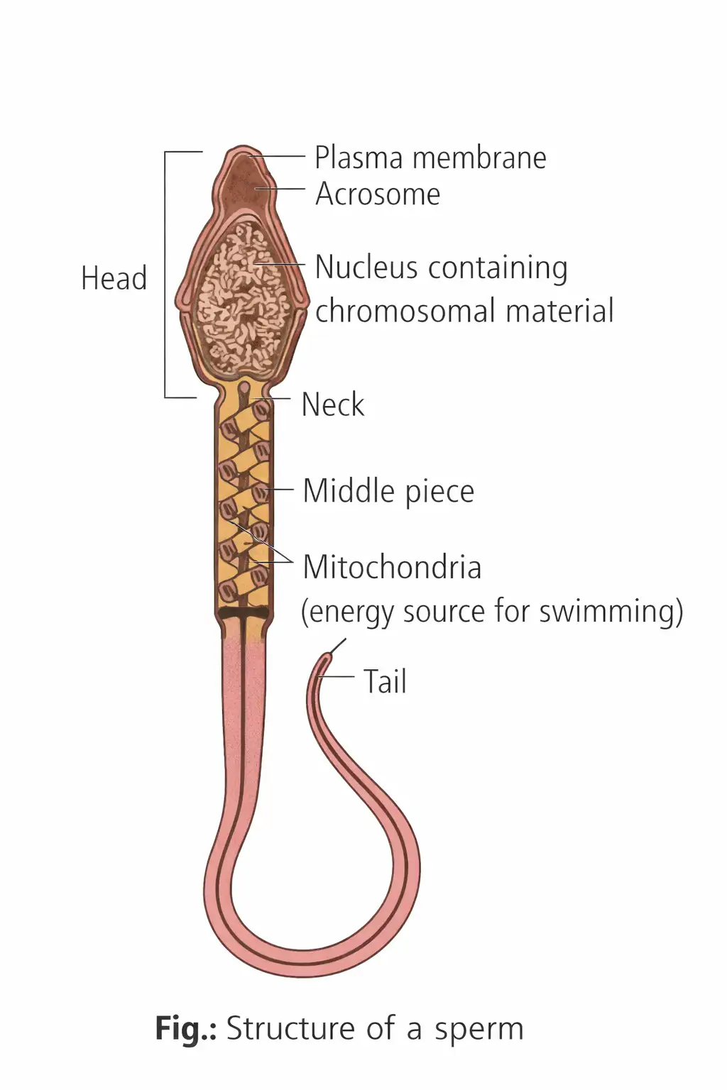
Spermiogenesis — The transformation of spermatids into spermatozoa (sperm); it is also called spermateliosis.
| Part | Structure or function |
|---|---|
| Head | Contains an elongated haploid nucleus; its front is covered by the enzyme-filled acrosome, which aids fertilisation. |
| Neck | One of the four named regions of the sperm. |
| Middle piece | Contains many mitochondria that supply energy for tail movement. |
| Tail | Enables sperm movement; its microtubular arrangement is \(9+2\). |
| Whole cell | The entire sperm is enclosed by a plasma membrane. |
Spermiation — The release of spermatozoa from Sertoli cells into the seminiferous tubules after spermiogenesis.
Sperm transport and semen
- Sperm output: A normal ejaculation releases about \(200\)–\(300\) million sperms; high production helps offset the odds against internal fertilisation.
- Transport and maturation: Accessory ducts carry sperm; secretions from the epididymis, vas deferens, seminal vesicles and prostate support sperm maturation and motility.
- Semen: A mixture of sperm and seminal plasma.
- Hormonal support: Androgens maintain the functions of male accessory ducts and glands.
Spermatocytogenesis — The formation of primary spermatocytes from spermatogonia.
Hormonal Control in Males
Hormonal regulation of spermatogenesis
- At puberty, increased hypothalamic GnRH stimulates the anterior pituitary.
- The anterior pituitary releases LH and FSH.
- LH stimulates Leydig cells to secrete testosterone; FSH stimulates Sertoli cells to secrete androgen-binding protein (ABP) and inhibin.
- Testosterone inhibits LH secretion, while inhibin inhibits FSH secretion by negative feedback.
Oogenesis
Oogenesis — The process by which a mature female gamete (ovum) is formed.
Oogenesis begins before birth
- Oogonia: About a couple of million gamete mother cells form in each fetal ovary; no new oogonia are added after birth.
- Arrest: Oogonia enter meiosis and primary oocytes arrest in prophase I at the diplotene stage.
- Primary follicle: Each primary oocyte becomes surrounded by a layer of granulosa cells.
- Follicle loss: Many follicles degenerate from birth to puberty; about \(60{,}000\)–\(80{,}000\) primary follicles remain in each ovary at puberty.
Follicle development and oocyte maturation
- Primary follicle develops additional granulosa layers and a theca, becoming a secondary follicle.
- The secondary follicle becomes a tertiary follicle with a fluid-filled cavity called the antrum.
- The primary oocyte grows and completes meiosis I unequally, producing a large haploid secondary oocyte and a small first polar body.
- The follicle matures into a Graafian follicle; the secondary oocyte is surrounded by the zona pellucida.
- Ovulation releases the secondary oocyte from the ruptured Graafian follicle.
- Usually after sperm entry in the oviduct, the secondary oocyte completes meiosis II, forming a large ootid and a small second polar body; the ootid develops into the ovum.
Oogenesis outcomes
- Unequal divisions: Most cytoplasm remains in the functional ovum; the polar bodies receive little cytoplasm.
- Final products: One oogonium ultimately forms one haploid ovum and three polar bodies; the first polar body may divide to form two.
- At ovulation: Humans release a secondary oocyte, not a fully mature ovum; meiosis II is completed after sperm entry.
- Mature ovum: Spherical, contains a nucleus and is the largest cell in the body.
Vitellogenesis — The deposition of yolk in the primary oocyte during prophase I.
Hormonal Control in Females
Hormonal regulation of oogenesis
- Hypothalamic GnRH stimulates the anterior pituitary to release LH and FSH.
- FSH promotes ovarian follicle growth, oocyte development and completion of meiosis I; it also stimulates oestrogen production.
- LH stimulates the corpus luteum to secrete progesterone.
- Rising progesterone inhibits GnRH release, which in turn reduces FSH, LH and progesterone production.
What is the difference between spermiogenesis and spermiation?
What are the products of one primary spermatocyte and one oogonium?
Which cell is released at human ovulation, and when is meiosis II completed?
Quick recap
- Spermatogenesis begins at puberty in seminiferous tubules and produces four haploid spermatids per primary spermatocyte.
- Spermiogenesis forms spermatozoa; spermiation releases them from Sertoli cells.
- GnRH stimulates pituitary LH and FSH; LH acts on Leydig cells, while FSH acts on Sertoli cells.
- Oogenesis starts before birth; primary oocytes arrest in prophase I at diplotene.
- Meiosis I produces a secondary oocyte and first polar body; ovulation releases the secondary oocyte.
- Meiosis II is usually completed after sperm entry, yielding one ovum and three polar bodies per oogonium.
- FSH supports follicular growth and oestrogen secretion; LH stimulates corpus luteum progesterone secretion, whose rise feeds back to reduce GnRH, FSH, LH and progesterone.
Menstrual Cycle
Overview
Menstrual Phase
Follicular or Proliferative Phase
Ovulatory Phase
Luteal or Secretory Phase
Estrous Cycle
What causes ovulation in the menstrual cycle?
What happens to the corpus luteum if fertilisation does not occur?
Quick recap
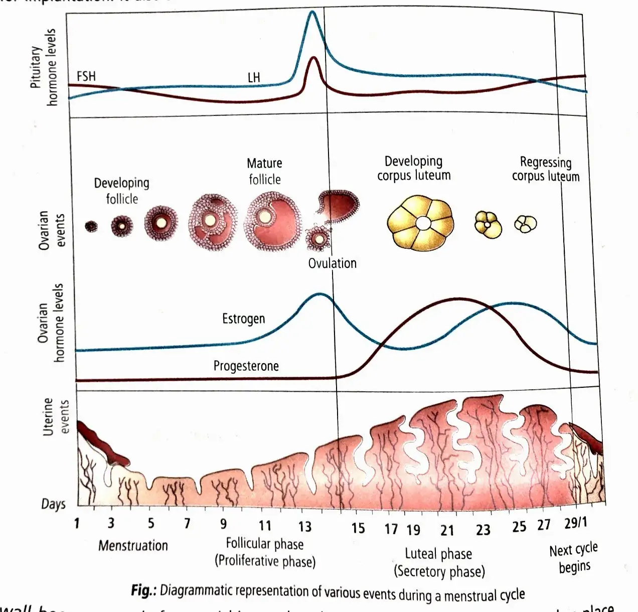
- The menstrual cycle in human females averages about 28–29 days; day 1 is the first day of menstruation.
- Menstrual bleeding follows the fall in progesterone and estrogen and the shedding of the endometrium.
- During the follicular phase, FSH supports follicle growth and estrogen promotes endometrial proliferation.
- The mid-cycle LH surge triggers ovulation, usually around day 14.
- The corpus luteum secretes progesterone to prepare and maintain the endometrium for implantation.
- Without fertilisation, corpus luteum degeneration leads to menstruation; pregnancy stops the cycle.
- Menarche begins cyclic menstruation; menopause marks its cessation, usually around age 50.
Fertilisation and Implantation
Insemination and Transport of Gametes
Insemination — The release of semen by the penis into the vagina during copulation.
Gamete transport
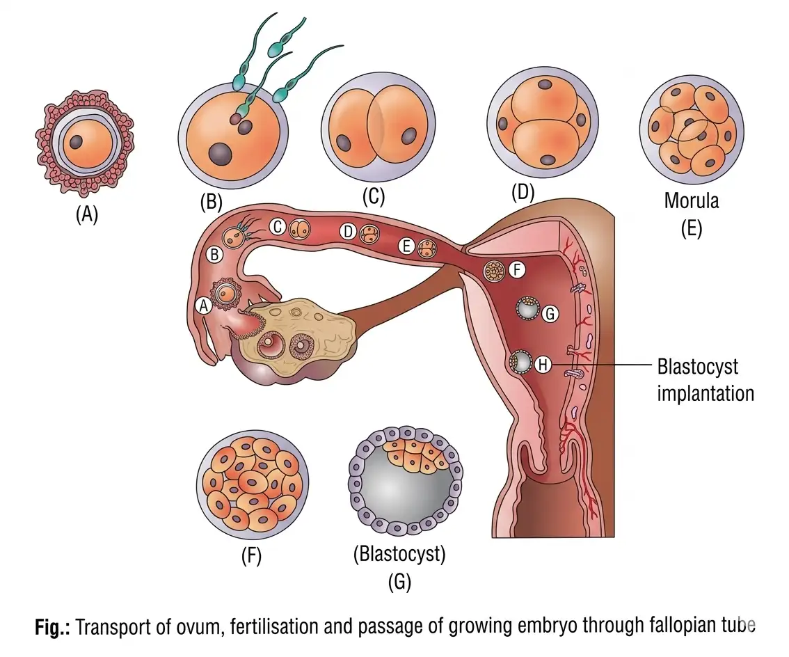
- Oocyte route: The secondary oocyte released at ovulation is received by the fallopian tube; ciliated cells in the infundibulum move it towards the ampulla.
- Fertilisation site: The ampullary–isthmic junction; fertilisation requires the ovum and sperms to reach the ampullary region simultaneously.
- Sperm movement: Uterine and fallopian-tube contractions assist sperm initially; sperm then move by their own motility at about 1.5–3 mm/min.
- Timing: The secondary oocyte can be fertilised for only about 24 hours after ovulation; otherwise, it degenerates.
Capacitation and Acrosomal Reaction
Capacitation — Activation of sperm in the female reproductive tract, enabling it to fertilise the egg.
Sperm activation
- Female tract secretions: Remove coating substances, especially over the acrosome, exposing sperm receptor sites; capacitation takes about 5–6 hours.
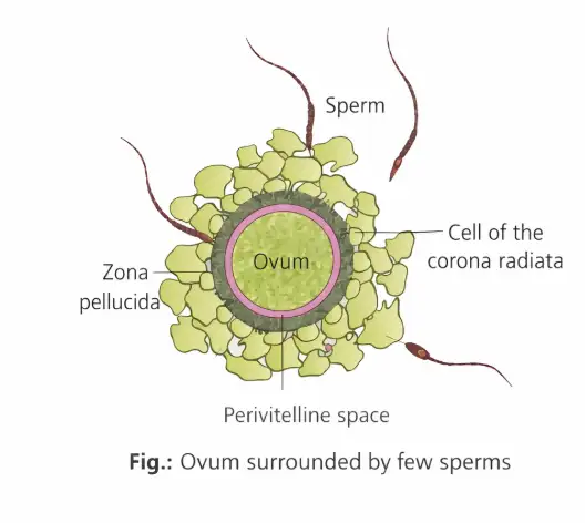
- Acrosomal reaction: The capacitated sperm releases sperm lysins from its acrosome to penetrate the egg coverings.
- Membrane fusion: During fertilisation, the acrosomal plasma membrane fuses with the oocyte plasma membrane.
| Lysin | Action |
|---|---|
| Hyaluronidase | Acts on the ground substance of follicle cells. |
| Corona-penetrating enzymes | Dissolve the corona radiata. |
| Acrosin | Digests the zona pellucida. |
Blocking entry of additional sperms
- Cortical reaction: Sperm contact with the zona pellucida triggers changes in the oocyte membrane.
- Result: These changes prevent additional sperms from entering, ensuring that only one sperm fertilises the ovum.
Formation of the Zygote and Fertilisation Theory
Formation of the diploid zygote
- Sperm enters the oocyte through the zona pellucida and plasma membrane.
- Sperm entry induces completion of the secondary oocyte’s second meiotic division.
- Unequal division produces a second polar body and a haploid ovum (ootid).
- The haploid sperm and ovum nuclei fuse to form a diploid zygote.
Fertilizin theory
- Proposal: In 1914, F. R. Lillie proposed that the ovum secretes fertilizin and sperm carry antifertilizin on their surface.
- Composition: Fertilizin is a glycoprotein made of monosaccharides and amino acids; antifertilizin consists of acidic amino acids.
- Species recognition: Matching fertilizin and antifertilizin bind sperm to the egg of the same species.
- Agglutination: This chemical recognition and adhesion of sperm to an egg of the same species is called agglutination.
Sex Determination and Cleavage
| Gamete / zygote | Sex chromosome(s) | Outcome |
|---|---|---|
| Female | \(X\) in every haploid gamete | The ovum contributes \(X\). |
| Male | \(X\) or \(Y\); about \(50\%\) of sperms carry each | The sperm determines whether the zygote is \(XX\) or \(XY\). |
| Zygote \(XX\) | \(XX\) | Develops as a female. |
| Zygote \(XY\) | \(XY\) | Develops as a male. |
Cleavage — Rapid, repeated mitotic divisions of the zygote that increase cell number without increasing the embryo’s overall volume.
Cleavage and early development
- Location: Cleavage begins as the zygote moves through the isthmus of the oviduct towards the uterus.
- Cell stages: The zygote divides into 2, 4, 8 and then 16 blastomeres.
- Morula: The embryo with 8–16 blastomeres is called a morula.
- Blastocyst: Continued division forms a blastocyst with an outer trophoblast and an inner cell mass attached to it.
- Cell fate: The trophoblast attaches to the endometrium; the inner cell mass differentiates to form the embryo.
| Function | Significance |
|---|---|
| Distributes zygote cytoplasm | Partitions the cytoplasm among blastomeres. |
| Increases protoplasmic mobility | Supports morphogenetic movements for differentiation, germ-layer formation and formation of tissues and organs. |
| Restores cell size and nucleocytoplasmic ratio | Establishes values characteristic of the species. |
| Builds cell number | Converts the unicellular zygote into a multicellular embryo. |
Blastocyst cell groups
- Holoblastic cleavage: Human blastomeres divide completely.
- Cells of Rauber: Trophoblast cells in contact with the inner cell mass; they later contribute to the chorion, amnion and parts of the placenta.
- Trophoblast role: It remains external to the embryo and provides nutrition; it does not form the embryo.
- Inner cell mass: The precursor of the embryo.
Implantation
Implantation — Embedding of the blastocyst in the uterine endometrium; this leads to pregnancy.
Implantation sequence
- Timing: Implantation occurs about the seventh day after fertilisation.
- Orientation: The blastocyst side containing the inner cell mass lies against the uterine endometrium.
- Zona hatching: The blastocyst sheds its zona pellucida before implantation.
- Attachment and embedding: Trophoblast attaches to the endometrium; uterine cells divide rapidly and cover the blastocyst, embedding it in the endometrium.
- Zona pellucida role: It prevents abnormal-site implantation by keeping sticky, phagocytic trophoblast cells from being exposed until the blastocyst reaches the proper site.
What is capacitation, and how does it help fertilisation?
What are the cell groups of a blastocyst, and what do they form?
What is zona hatching, and when does implantation occur?
Quick recap
- Insemination deposits semen in the vagina; fertilisation normally occurs at the ampullary–isthmic junction.
- Capacitation activates sperm; acrosomal lysins help it penetrate egg coverings, and the cortical reaction blocks polyspermy.
- Sperm entry completes meiosis II; fusion of the haploid nuclei forms the diploid zygote.
- The sperm contributes either \(X\) or \(Y\), producing an \(XX\) or \(XY\) zygote.
- Cleavage forms blastomeres, then a morula and a blastocyst with trophoblast and inner cell mass.
- After zona hatching, the blastocyst implants in the endometrium around day seven after fertilisation.
Pregnancy and Embryonic Development
Implantation and Placenta
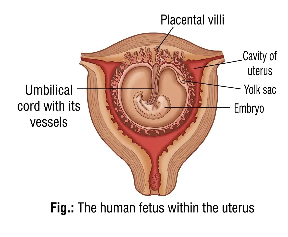
Placenta — A structural and functional unit formed by interdigitation of chorionic villi and uterine tissue, linking the developing embryo or fetus with the mother.
Placental structure and functions
- Formation: After implantation, finger-like chorionic villi develop from the trophoblast; uterine tissue and maternal blood surround them.
- Interdigitation: Chorionic villi and uterine tissue interlock to form the placenta.
- Exchange: The placenta supplies oxygen and nutrients to the embryo and removes carbon dioxide and embryonic wastes.
- Connection: The umbilical cord transports substances to and from the embryo.
- Endocrine role: The placenta secretes hCG, hPL, estrogens and progestogens.
Pregnancy Hormones
| Hormone(s) | Source or change | Key point |
|---|---|---|
| hCG, hPL, estrogens and progestogens | Secreted by the placenta | hCG and hPL are produced only during pregnancy |
| Relaxin | Secreted by the ovary in the later stage of pregnancy | Produced only during pregnancy |
| Estrogens, progestogens, cortisol, prolactin and thyroxine | Maternal blood levels rise several-fold during pregnancy | Higher levels support fetal growth, maternal metabolic changes and maintenance of pregnancy |
Early Embryonic Structures and Membranes
Blastocyst differentiation
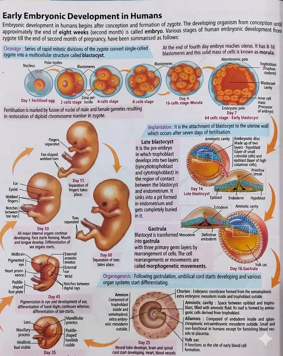
- About 8 days after fertilisation, the trophoblast develops into two layers: syncytiotrophoblast and cytotrophoblast.
- The inner cell mass contains stem cells capable of giving rise to all tissues and organs.
- In the late blastocyst, the inner cell mass differentiates into epiblast and hypoblast; together they form the embryonic disc.
- Differentiation between the epiblast and trophoblast generates the amniotic cavity, which is filled with amniotic fluid.
| Structure | Formation | Key feature |
|---|---|---|
| Extra-embryonic mesoderm | Trophoblast cells give rise to a mass that differentiates into outer somatopleuric and inner splanchnopleuric layers | The two layers enclose the extra-embryonic coelom |
| Chorion | Trophoblast outside and somatopleuric extra-embryonic mesoderm inside | Later becomes the main embryonic part of the placenta and produces hCG |
| Amnion | Amniogenic cells inside and somatopleuric extra-embryonic membrane outside | The amniotic cavity contains fluid that acts as a shock absorber |
| Yolk sac | Flattened hypoblast cells line the primary yolk sac; it becomes smaller after extra-embryonic mesoderm and coelom appear | The smaller later form is the secondary yolk sac |
Gastrulation — Transformation of the blastocyst into a gastrula through cell rearrangement, forming the primary germ layers.
Germ layers across animals
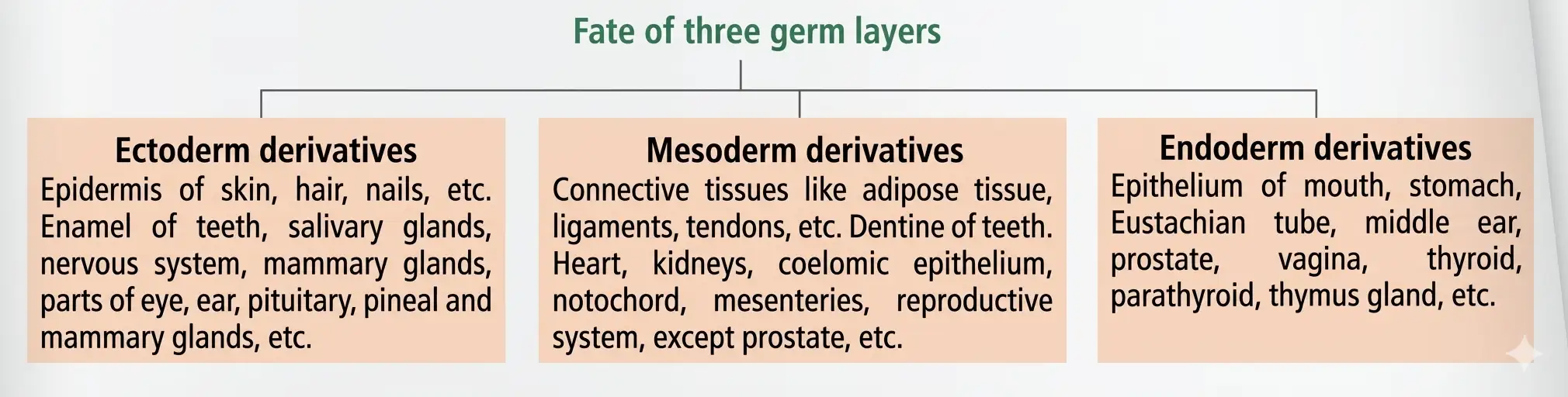
- Formation: In triploblastic animals, ectoderm, mesoderm and endoderm form through characteristic movements of some cells.
- Fate: The germ layers have similar fates across animals and give rise to specific tissues, organs and organ systems.
Primitive Streak and Formation of Germ Layers
Primitive streak
- Appearance: A faint groove forms on the dorsal surface of the epiblast during gastrulation and elongates from the posterior along the embryo.
- Axis formation: It establishes the embryo’s head–tail ends and right–left sides.
- Invagination: Epiblast cells move inward beneath the primitive streak and detach from the epiblast.
Formation of the three germ layers
- Some invaginating epiblast cells displace the hypoblast → endoderm forms.
- Other invaginating cells remain between the epiblast and new endoderm → mesoderm forms.
- Cells remaining in the epiblast → ectoderm forms.
- Ectoderm, mesoderm and endoderm → give rise to the body’s tissues, organs and organ systems.
How is the placenta formed, and what are its main functions?
How are the three primary germ layers formed during gastrulation?
Quick recap
- Implantation leads to pregnancy; chorionic villi interdigitate with uterine tissue to form the placenta.
- The placenta exchanges substances, connects to the embryo through the umbilical cord and secretes hormones.
- hCG, hPL and relaxin are produced only during pregnancy; several other maternal hormone levels rise several-fold.
- About 8 days after fertilisation, the trophoblast differentiates into syncytiotrophoblast and cytotrophoblast.
- The epiblast and hypoblast form the embryonic disc; extra-embryonic tissues form the chorion, amnion and yolk sac.
- The amniotic cavity is filled with fluid that cushions the embryo; the primitive streak establishes body axes and permits epiblast invagination.
- Gastrulation forms ectoderm, mesoderm and endoderm through characteristic cell movements; their fates are similar across animals.
Parturition and Lactation
Parturition
Lactation
Quick recap
- Parturition is the expulsion of the full-term baby at the end of gestation.
- Fetal and placental signals initiate the fetal ejection reflex, stimulating maternal oxytocin release.
- Oxytocin strengthens uterine contractions; relaxin increases pelvic flexibility and helps dilate the cervix.
- Labour is the forceful uterine contraction process that leads to birth.
- Afterbirth refers to expulsion of the placenta and remaining umbilical cord after birth.
- Colostrum is produced during the first 2–3 days of lactation and provides antibodies to the newborn.
NEET Zoology — ALL CHAPTERS
1
Structural Organisation in Animals
2
Breathing and Exchange of Gases
3
Body Fluids and Circulation
4
Excretory Products and their Elimination
5
Locomotion and Movement
6
Neural Control and Coordination
7
Human Reproduction
8
Reproductive Health
9
Evolution
10
Microbes in Human Welfare
11
Organism and Population
12
Ecosystem
13
Biodiversity and Conservation