NEET Ug . NEET Zoology . Zoology . Body Fluids and Circulation
Chapter 3: Body Fluids and Circulation
Ch 3
NEET Zoology
NEET Ug
Blood
Blood and Its Components
Blood — Blood is a mobile connective tissue made of a fluid matrix (plasma) and formed elements.
Role and Composition
- Transport: Blood carries nutrients, \(O_2\) and other essential substances to cells, and removes wastes; lymph also transports certain substances.
- Volume: Adult men have about 5–5.5 L of blood, constituting 30–35% of extracellular fluid.
- pH: Blood is slightly alkaline, with a pH of about 7.4.
- Components: Plasma forms about 55% of blood; formed elements form nearly 45%.
Plasma
Plasma — Plasma is the pale-yellow, viscous, slightly alkaline, non-living fluid matrix of blood.
- Composition: About 90–92% water and 6–8% plasma proteins; the rest includes dissolved substances.
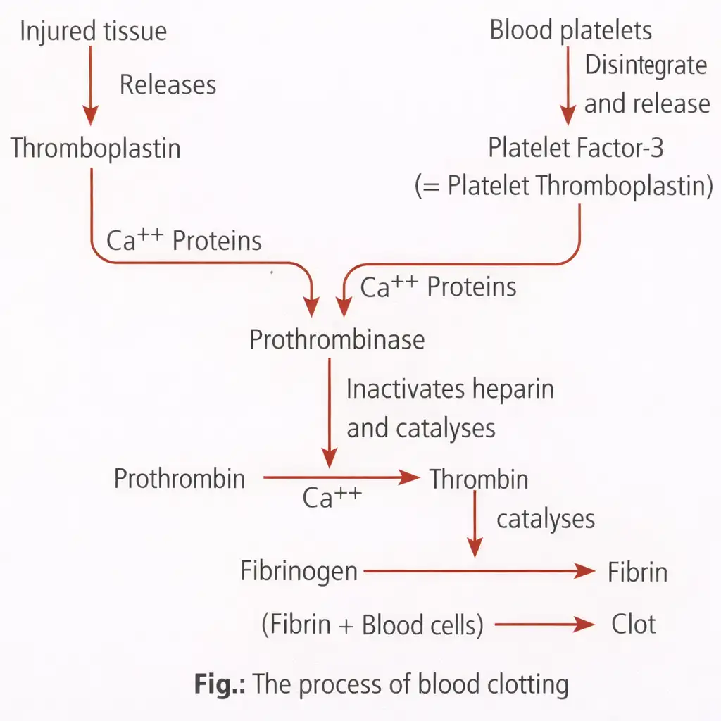
- Proteins: Albumin helps maintain osmotic balance; globulins and immunoglobulins aid defence; fibrinogen and prothrombin are involved in clotting.
- Other contents: Minerals such as \(Na^+\), \(Ca^{2+}\), \(Mg^{2+}\), \(HCO_3^-\) and \(Cl^-\), plus glucose, amino acids and lipids in transit.
- Clotting factors: Present in plasma in inactive forms.
Serum — Serum is plasma without clotting factors.
Formed Elements
Formed elements — Erythrocytes, leucocytes and platelets are the cellular components of blood and make up nearly 45% of it.
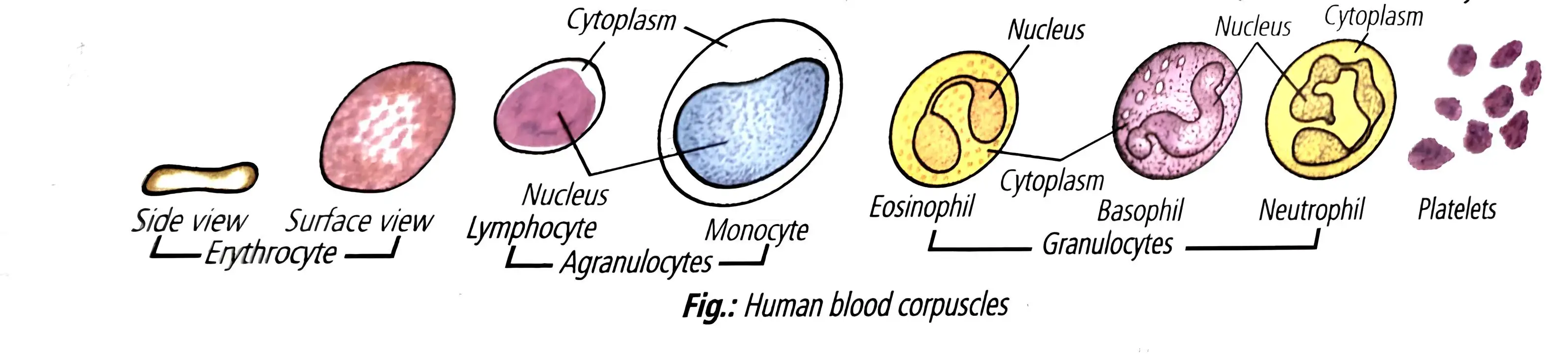
Erythrocytes (Red Blood Cells)
Erythrocytes — Erythrocytes, or red blood cells (RBCs), are the most abundant blood cells and carry haemoglobin.
- Count: About 5 million RBCs per \(mm^3\) in adult men and 5.5 million per \(mm^3\) in adult women.
- Formation: Produced in adult red bone marrow; RBC formation is called erythropoiesis.
- Structure: Mature mammalian RBCs are circular, biconcave and enucleated. Their shape provides a large surface area for gaseous exchange.
- Haemoglobin: Gives blood its red colour; a healthy individual has about 12–16 g per 100 mL of blood.
- Requirements: Iron, proteins, vitamin \(B_{12}\) and folic acid are needed for RBC and haemoglobin formation.
- Life and destruction: RBCs live about 120 days and are destroyed in the spleen, the “graveyard of RBCs.”
- Altitude: People living in hills have more RBCs to cope with the lower oxygen content of the air.
| Condition | Meaning / effect |
|---|---|
| Polycythemia | Abnormal increase in RBC count. |
| Erythrocytopenia | Decrease in RBC count; can cause oxygen shortage in blood and tissues. |
| Anaemia | Low haemoglobin level. |
Leucocytes (White Blood Cells)
Leucocytes — Leucocytes, or white blood cells (WBCs), are colourless, nucleated blood cells that protect the body.
- Count and lifespan: About 6,000–8,000 per \(mm^3\) of blood; WBCs are generally short-lived.
- Classes: Granulocytes are neutrophils, eosinophils and basophils; agranulocytes are lymphocytes and monocytes.
- Extremes: Neutrophils are the most numerous WBCs; monocytes are the largest.
- Formation and disorders: WBC formation is leucopoiesis; a decreased count is leukopenia, while abnormal increases occur in malignancies such as leukaemia.
| Type | Class | Proportion | Nucleus | Key function |
|---|---|---|---|---|
| Neutrophils | Granulocyte | 60–65% | Many-lobed | Phagocytose harmful germs and foreign particles. |
| Basophils | Granulocyte | 0–1% | Three-lobed, S-shaped | Release heparin, serotonin and histamine involved in inflammatory reactions. |
| Eosinophils | Granulocyte | 2–3% | Bilobed | Numbers increase in allergic conditions such as asthma and hay fever. |
| Lymphocytes | Agranulocyte | 20–25% | Large, rounded | Produce antibodies; B and T lymphocytes mediate immune responses. |
| Monocytes | Agranulocyte | 2–10% | Bean-shaped | Phagocytose bacteria and cellular debris. |
Platelets
Platelets — Platelets, or thrombocytes, are small cell fragments that help blood clot.
- Origin and form: Produced from bone-marrow megakaryocytes; they are colourless, rounded or oval disc-like fragments.
- Count: About 150,000–350,000 per \(mm^3\) of blood.
- Function: Release substances involved in blood clotting.
- Life and low count: Their lifespan is about one week; a reduced count can cause clotting disorders and excessive blood loss.
Blood Groups
- Basis: Blood groups depend on antigens on RBCs and antibodies in plasma.
- Transfusion safety: Donor and recipient groups must be matched to avoid clumping and destruction of RBCs.
ABO Blood Group System
- Discovery: Karl Landsteiner reported the ABO groups in 1900; Decastello and Sturli identified the AB group in 1902.
- Genetic control: Gene \(I\) has three alleles: \(I^A\), \(I^B\) and \(I^O\).
- Antigens: Alleles \(I^A\) and \(I^B\) produce A and B antigens, respectively, on RBC surfaces.
- Antibodies: Plasma contains antibodies against the A or B antigen absent from a person's RBCs.
| Blood group | Genotype | RBC antigen(s) | Plasma antibody(ies) | Can donate to | Can receive from |
|---|---|---|---|---|---|
| A | \(I^A I^A\) or \(I^A I^O\) | A | Anti-B | A, AB | A, O |
| B | \(I^B I^B\) or \(I^B I^O\) | B | Anti-A | B, AB | B, O |
| AB | \(I^A I^B\) | A and B | None | AB | A, B, AB, O |
| O | \(I^O I^O\) | None | Anti-A and Anti-B | A, B, AB, O | O |
- ABO red-cell transfusion: O is the universal donor and AB the universal recipient.
Rh Blood Group System
- Discovery: Landsteiner and Wiener identified the Rh antigen in 1940 in the blood of a Rhesus monkey.
- Types: Nearly 80% of people have the antigen and are Rh-positive (\(Rh^+\)); those without it are Rh-negative (\(Rh^-\)).
- Antibodies: An \(Rh^-\) person exposed to \(Rh^+\) blood can form antibodies against the Rh antigen; Rh status must also be matched for transfusions.
Rh Incompatibility in Pregnancy
Erythroblastosis fetalis — Erythroblastosis fetalis is the destruction of fetal RBCs when maternal Rh antibodies enter the blood of an Rh-positive fetus.
Rh Sensitisation and Its Consequences
- An \(Rh^-\) mother carries an \(Rh^+\) fetus; their blood is usually separated by the placenta.
- At the first delivery, fetal blood may enter the mother and stimulate production of anti-Rh antibodies.
- In a later \(Rh^+\) pregnancy, maternal antibodies can cross the placenta and destroy fetal RBCs.
- The result may be severe anaemia, jaundice or fetal death; anti-Rh antibodies given to the mother immediately after the first delivery can prevent the condition.
Blood Coagulation
Blood coagulation — Blood coagulation is the process that stops bleeding after vessel injury and prevents excessive blood loss.
- Clot: A dark reddish-brown mass formed at the injury site; a fibrin-thread network traps dead and damaged formed elements.
- Clotting time: Usually 3–8 minutes.
- Vitamin K: Essential because it is needed for prothrombin synthesis in the liver.
- Anticoagulants: Heparin and sodium or potassium oxalate prevent blood clotting.
Differentiate between plasma and serum.
What is erythroblastosis fetalis, and how can it be prevented?
State the roles of albumin, globulins and fibrinogen in plasma.
Quick recap
- Blood is a mobile connective tissue: plasma forms about 55% and formed elements about 45%; lymph also transports certain substances.
- Plasma contains water, proteins, dissolved nutrients, minerals and inactive clotting factors; serum lacks clotting factors.
- RBCs carry haemoglobin, WBCs defend the body, and platelets help clot blood.
- ABO compatibility depends on RBC antigens and plasma antibodies; O is the universal ABO donor and AB the universal recipient for red-cell transfusions.
- Rh incompatibility can cause erythroblastosis fetalis in a later pregnancy; anti-Rh antibodies can prevent it.
- Fibrin forms the clot network; vitamin K supports prothrombin synthesis, while anticoagulants prevent clotting.
Lymph (Tissue Fluid)
Lymph: Composition and Collection
Lymph — A mobile connective tissue made up of lymph plasma and lymph corpuscles.
Composition and Collection
- Lymph plasma: Resembles blood plasma but has fewer proteins, less calcium and phosphorus, and a higher glucose concentration.
- Lymph corpuscles: Floating amoeboid leucocytes, mostly lymphocytes.
- Absent: Erythrocytes and platelets are not present in lymph.
- Collection: The lymphatic system collects lymph and drains it back into the major veins.
Functions of Lymph
- Transport: Carries nutrients and hormones to body cells and returns carbon dioxide and other metabolic wastes from cells to the blood; these ultimately enter the venous system.
- Fat absorption: Fats are absorbed through lymph in the lacteals of the intestinal villi.
- Defence: Lymph nodes destroy invading microorganisms and foreign particles.
Quick recap
- Lymph is mobile connective tissue containing lymph plasma and lymph corpuscles.
- Lymph plasma has fewer proteins, less calcium and phosphorus, and more glucose than blood plasma.
- Lymph corpuscles are mostly lymphocytes; erythrocytes and platelets are absent.
- The lymphatic system collects lymph and drains it into major veins.
- Lymph transports nutrients and hormones to cells and carries carbon dioxide and other wastes from cells to blood, then to the venous system.
- Fats are absorbed through lymph in lacteals of intestinal villi; lymph nodes destroy microorganisms and foreign particles.
Circulatory Pathways
Circulatory Pathways and Vertebrate Hearts
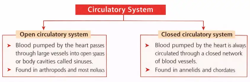
| Point | Open system | Closed system |
|---|---|---|
| Fluid pathway | Circulatory fluid leaves vessels and directly bathes tissues. | Blood remains within a closed network of vessels. |
| Control of flow | Flow is less precisely regulated. | Flow can be regulated more precisely; this is an advantage. |
| Heart type | Chambers and circulation | Groups | Blood flow |
|---|---|---|---|
| Two-chambered | One atrium and one ventricle; single circulation. | Fishes | The heart pumps deoxygenated blood to the gills; oxygenated blood goes to the body and returns deoxygenated to the heart. |
| Three-chambered | Two atria and one ventricle; incomplete double circulation. | Amphibians and reptiles | Oxygenated and deoxygenated blood enter separate atria but mix in the single ventricle. |
| Four-chambered | Two atria and two ventricles; double circulation. | Crocodiles, birds and mammals | Oxygenated and deoxygenated blood remain separate and are pumped through distinct pathways. |
Human Circulatory System
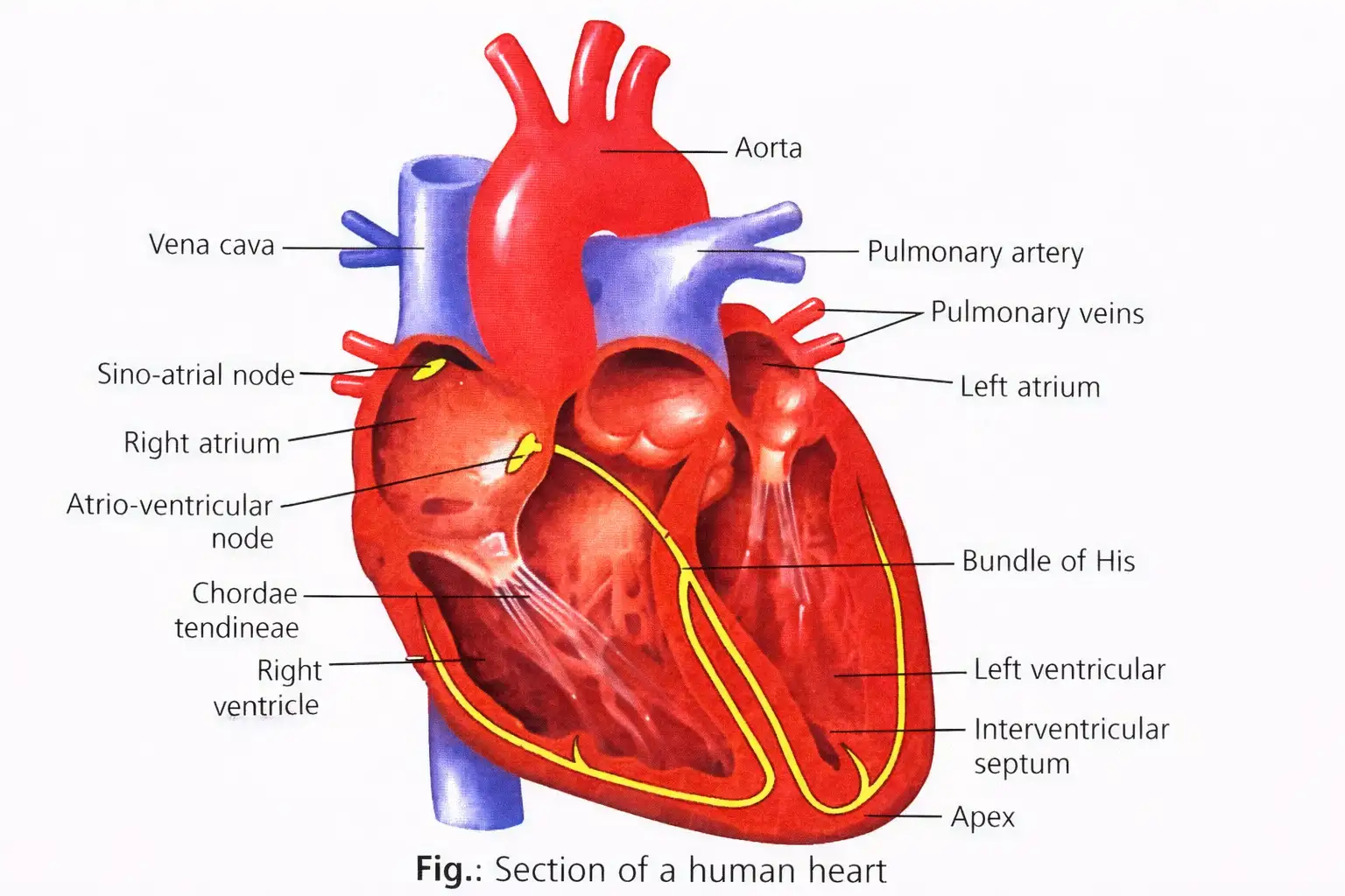
Human circulatory system — The blood vascular system consists of a muscular, chambered heart, blood and a closed network of branching blood vessels.
Heart: Structure and Major Vessels
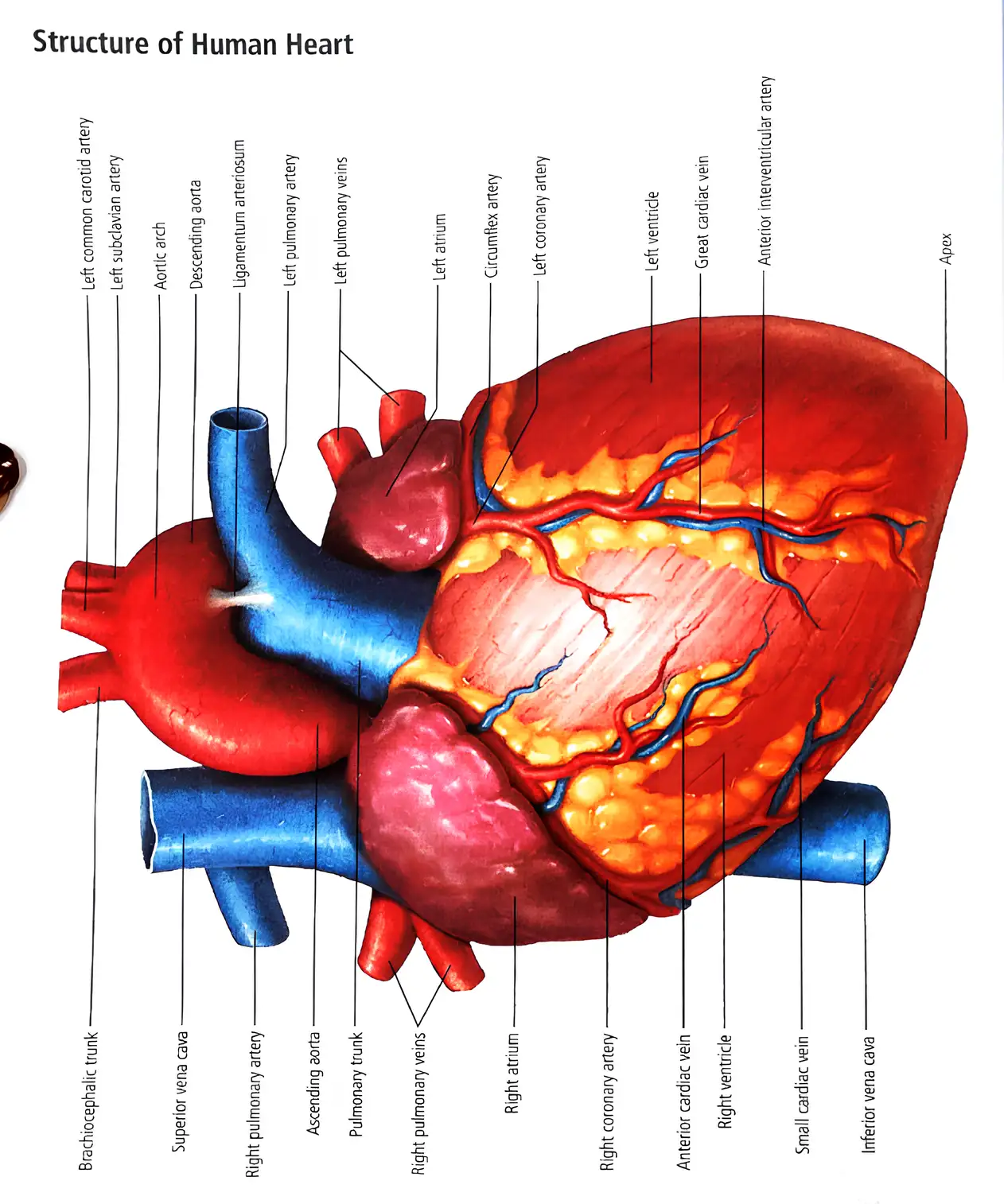
- Position and covering: The mesoderm-derived heart lies in the thoracic cavity between the lungs, slightly left of centre; it is about the size of a clenched fist.
- Pericardium: The four-chambered heart is enclosed in a pericardium. Pericardial fluid in the cavity between its two layers reduces friction.
- Chambers and septa: Right and left atria are separated by the interatrial septum; the ventricles are separated by the interventricular septum.
- Atrioventricular septum: Thick fibrous tissue separates each atrium from the ventricle on the same side.
- Right atrium: Receives deoxygenated blood from the superior vena cava (head and upper body) and inferior vena cava (lower body).
- Left atrium: Receives oxygenated blood from the lungs through the pulmonary veins.
- Ventricles: The left ventricular wall is thicker than the right because it pumps blood to the whole body at higher pressure.
| Valve | Position | Structure / role |
|---|---|---|
| Tricuspid | Right atrium to right ventricle | Three cusps; prevents backflow into the right atrium. |
| Bicuspid (mitral) | Left atrium to left ventricle | Two cusps; prevents backflow into the left atrium. |
| Pulmonary semilunar | Exit of the right ventricle into the pulmonary artery | Prevents blood returning to the right ventricle. |
| Aortic semilunar | Exit of the left ventricle into the aorta | Prevents blood returning to the left ventricle. |
Valve Support and Function
- Atrioventricular valves: The tricuspid and bicuspid valves lie between the atria and ventricles.
- Chordae tendineae: Fibrous cords connect the flaps of these valves to papillary muscles.
- One-way flow: Heart valves prevent regurgitation and maintain unidirectional blood flow.
Human Double Circulation
- Right ventricle → pulmonary artery → lungs (blood becomes oxygenated) → pulmonary veins → left atrium: pulmonary circulation.
- Left ventricle → aorta → body tissues (blood delivers oxygen) → venae cavae → right atrium: systemic circulation.
Cardiac Conduction and Heartbeat
Conduction Pathway
- Sinoatrial node (SAN) in the right atrium near the superior vena cava generates the impulse.
- The impulse spreads across the atria and reaches the atrioventricular node (AVN).
- The atrioventricular bundle (AV bundle) carries it through the atrioventricular septum and divides into right and left bundle branches.
- The branches give rise to Purkinje fibres, which distribute the impulse through the ventricular muscle; the branches and Purkinje fibres together are called the bundle of His.
Nodal Tissue
- Autoexcitable tissue: Cardiac nodal tissue can generate action potentials without external stimulation.
- SAN—pacemaker: Generates the highest number of impulses, about \(70\)–\(75\) per minute, and initiates and maintains the rhythmic heartbeat.
- AVN—pacesetter: Rapidly transmits the excitation received from the SAN to the ventricles through the bundle of His.
| Point | Neurogenic | Myogenic |
|---|---|---|
| Initiation | A nerve impulse from a nearby nerve ganglion initiates the heartbeat. | A specialised patch of heart muscle initiates the heartbeat. |
| Examples | Some annelids and most arthropods. | Molluscs and vertebrates, including humans. |
Cardiac Cycle
Cardiac cycle — The cardiac cycle is the sequence of atrial and ventricular contraction (systole) and relaxation (diastole) that repeats in the heart.
| Stage | Main events |
|---|---|
| Joint diastole | All chambers relax. Blood enters the atria from the venae cavae and pulmonary veins, then flows through open AV valves into the ventricles; semilunar valves are closed. |
| Atrial systole | The SAN triggers atrial contraction, pushing additional blood into the ventricles and increasing ventricular filling by about 30%. |
| Ventricular systole | The impulse reaches ventricular muscle through the AV bundle and bundle of His. Rising ventricular pressure closes the AV valves, producing the first sound, lub; the semilunar valves open and blood is ejected. |
| Ventricular diastole | Ventricular relaxation lowers pressure; semilunar valves close, producing the second sound, dub. When ventricular pressure falls below atrial pressure, the AV valves open and filling resumes, returning the heart to joint diastole. |
| Event / sound | Duration or frequency |
|---|---|
| Atrial systole | \(0.1\) s |
| Ventricular systole | \(0.3\) s |
| Joint diastole | \(0.4\) s |
| Complete cardiac cycle | \(0.8\) s |
| First sound (lub) | \(0.15\) s; \(25\)–\(45\) Hz; AV valve closure |
| Second sound (dub) | About \(0.12\) s; about \(50\) Hz; semilunar valve closure |
Cardiac output is the volume of blood pumped by one ventricle per minute. Stroke volume is the volume ejected by one ventricle per beat.
Cardiac Output Example
- Example: At \(72\) beats/min and a stroke volume of \(0.08\) L/beat, multiplication gives \(72\times0.08=5.76\) L/min, or about \(5.8\) L/min.
- Source figure: The lecture states approximately \(5.5\) L/min for these values; this does not match the multiplication, so use \(5.76\) L/min as the calculated result and note the source discrepancy if required.
- Adjustment: The body can alter both heart rate and stroke volume; an athlete can have a higher cardiac output than an ordinary person.
Electrocardiogram (ECG)
Electrocardiogram (ECG) — An ECG is a graphic record of the electrical activity produced by excitation of the cardiac muscles; it is recorded using an electrocardiograph.
Recording and ECG Waves
- Standard recording: Three electrical leads are attached—one to each wrist and one to the left ankle—to monitor heart activity.
- P wave: Small upward wave showing atrial depolarisation, initiated by SAN activity; it precedes atrial contraction.
- QRS complex: Q is a small downward deflection, R a large upward deflection and S a downward deflection. The complex represents ventricular depolarisation; Q marks the beginning of ventricular systole.
- T wave: Dome-shaped wave showing ventricular repolarisation, associated with ventricular relaxation; its end marks the end of systole.
- Clinical use: Counting QRS complexes over a time interval gives heart rate. An abnormal ECG shape can indicate disease or another abnormality.
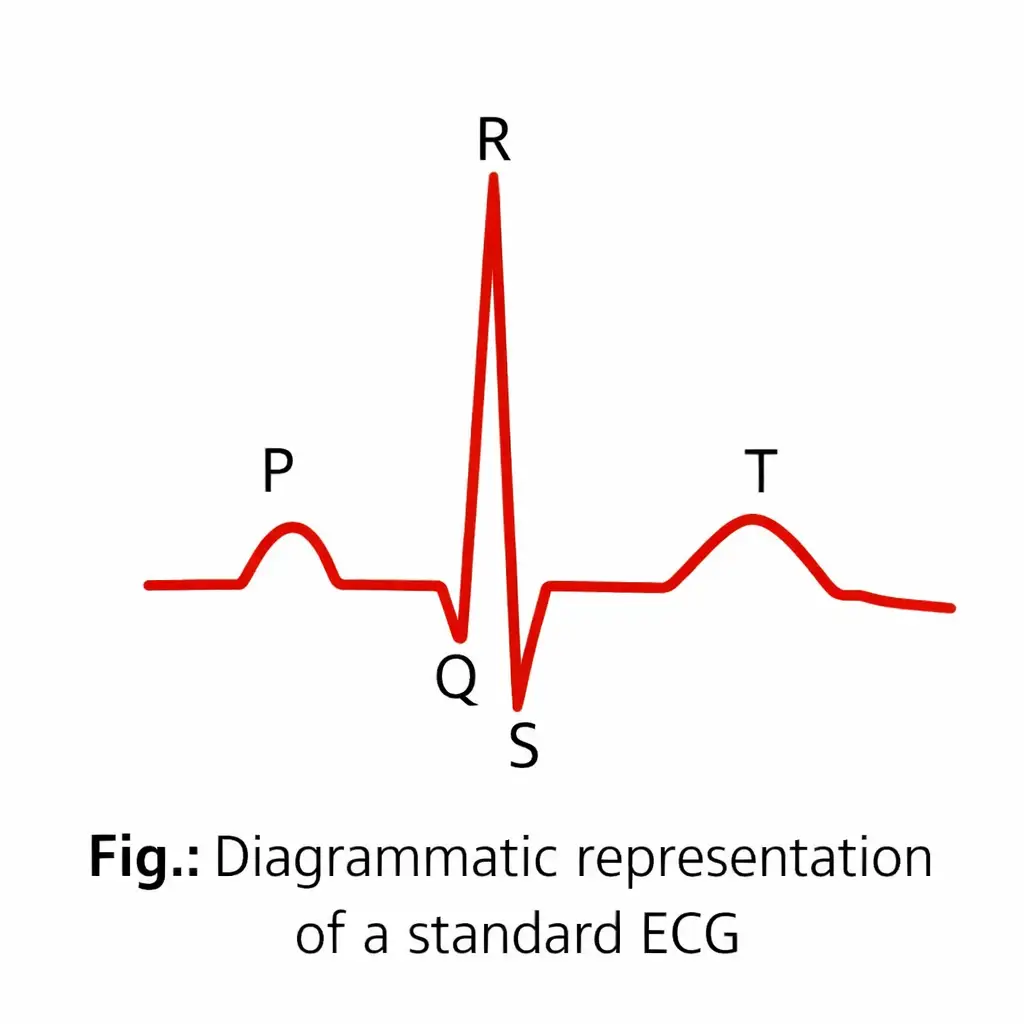
Do not confuse depolarisation with relaxation: the QRS complex represents ventricular depolarisation and initiates contraction; the T wave represents ventricular repolarisation and relaxation.
Why is the left ventricle thicker than the right ventricle?
What causes the first and second heart sounds?
Quick recap
- Closed circulation keeps blood within vessels and allows more precise regulation; vertebrate hearts range from two to four chambers.
- The human heart has four chambers, one-way valves and separate pulmonary and systemic pathways.
- The SAN initiates the heartbeat; impulses pass through the AVN, AV bundle, bundle branches and Purkinje fibres, collectively called the bundle of His.
- A cardiac cycle lasts \(0.8\) s; AV valve closure produces lub and semilunar valve closure produces dub.
- Cardiac output equals heart rate multiplied by stroke volume; \(72\) beats/min and \(0.08\) L/beat calculate to \(5.76\) L/min, despite the source's stated approximation of \(5.5\) L/min.
- P represents atrial depolarisation, QRS ventricular depolarisation, and T ventricular repolarisation.
Double Circulation
Blood Vessels
Blood vessels — Blood vessels transport blood through the body; the three types are arteries, veins and capillaries.
| Vessel | Direction and usual blood type | Key feature |
|---|---|---|
| Artery | Carries blood away from the heart; usually oxygenated | Elastic vessel; the pulmonary artery carries deoxygenated blood. |
| Vein | Carries blood towards the heart; usually deoxygenated | Valves prevent backflow; the pulmonary vein carries oxygenated blood from the lungs to the left atrium. |
| Capillary | Connects arteries and veins in tissues | Links the two types of vessels. |
Blood Vessel Wall Layers
| Layer | Position and composition |
|---|---|
| Tunica intima | Innermost; consists of squamous endothelium and an elastic membrane. |
| Tunica media | Middle; smooth involuntary muscle and elastic fibres. Comparatively thin in veins. |
| Tunica externa | Outermost; fibrous connective tissue containing collagen fibres. |
Double Circulation
Double circulation — The same blood passes through the heart twice along separate pathways during one complete cycle.
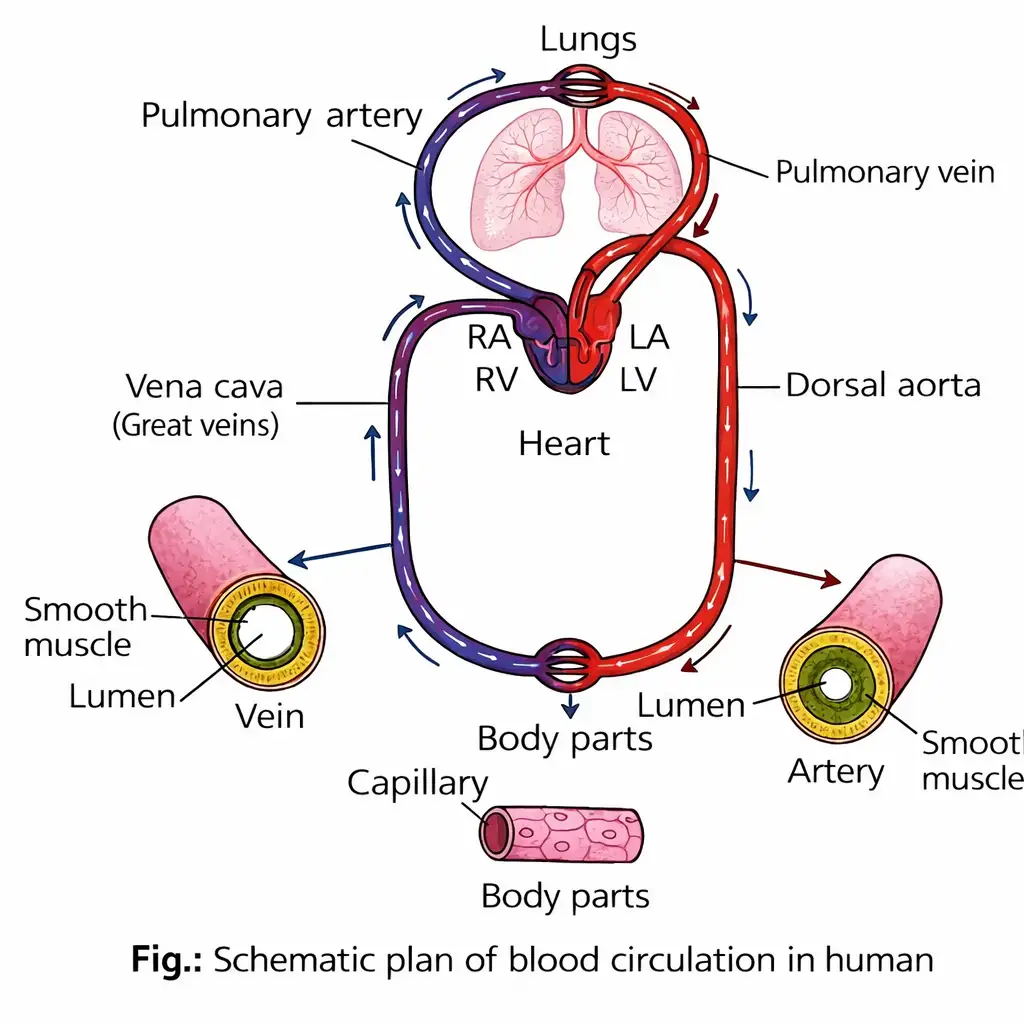
Pulmonary and Systemic Circulation
Pulmonary circulation — Blood moves from the heart to the lungs and back to the heart.
Systemic circulation — Oxygenated blood flows from the left ventricle to the body, and deoxygenated blood returns to the right atrium.
Special Circulations
Hepatic portal system — A special vascular connection between the digestive tract and the liver.
Coronary circulation — Blood circulation to and from the cardiac musculature through a special system of blood vessels.
Quick recap
- Arteries are elastic vessels that carry blood away from the heart; veins return blood, and capillaries link them in tissues.
- The pulmonary artery carries deoxygenated blood; pulmonary veins carry oxygenated blood to the left atrium.
- Artery and vein walls have tunica intima, tunica media and tunica externa; tunica media is thinner in veins.
- Double circulation has pulmonary and systemic circuits, so blood passes through the heart twice per cycle.
- Pulmonary route: right ventricle → lungs → left atrium; systemic route: left ventricle → body → right atrium.
- The hepatic portal system connects the digestive tract and liver; coronary circulation serves the cardiac musculature.
Regulation of Cardiac Activity
Intrinsic and Neural Regulation
Effects of Autonomic and Hormonal Signals
Quick recap
- The heart is myogenic because nodal tissue autoregulates its normal activity.
- The medulla oblongata moderates cardiac function through the ANS.
- Sympathetic signals increase heart rate and ventricular contraction strength, raising cardiac output.
- Parasympathetic signals reduce heart rate and action-potential conduction speed, lowering cardiac output.
- Adrenal medullary hormones can increase cardiac output.
Disorders of Circulatory System
Hypertension and Blood Pressure
Blood pressure and hypertension
- Hypertension: The most common disease affecting the heart and blood vessels; high blood pressure can harm the heart, brain, kidneys and eyes.
- Normal blood pressure: \(120/80\ \text{mmHg}\); the first value is systolic and the second is diastolic.
- Systolic pressure: Pressure produced when the left ventricle contracts and pushes blood into the aorta; normally about \(120\ \text{mmHg}\).
- Diastolic pressure: Pressure in the blood vessels when the heart is relaxed during complete diastole; normally about \(80\ \text{mmHg}\).
- Hypertension threshold: Blood pressure above \(140\ \text{mmHg}\) systolic and \(90\ \text{mmHg}\) diastolic.
- Measurement: A sphygmomanometer measures systolic and diastolic blood pressure.
| Measure | Meaning | Value |
|---|---|---|
| Systolic pressure | Pressure during ventricular contraction | \(120\ \text{mmHg}\) |
| Diastolic pressure | Pressure during heart relaxation | \(80\ \text{mmHg}\) |
| Pulse pressure | Difference between systolic and diastolic pressure | \(40\ \text{mmHg}\) for a normal reading |
Subtract diastolic pressure from systolic pressure to calculate pulse pressure.
Atherosclerosis and Arteriosclerosis
Atherosclerosis
- Plaque composition: Lipids such as cholesterol, fat, calcium and fibrous tissue collect on the inner lining of arteries.
- Effect: Plaques may completely block an artery; in coronary arteries, they reduce blood supply to the heart muscle.
- Consequence: Reduced coronary blood flow can lead to a heart attack or stroke.
Arteriosclerosis — Hardening and thickening of arteries due to deposits of calcium salts and cholesterol.
Arteriosclerosis
- Effect: The artery loses its ability to distend, and its walls may rupture.
- Risk: When coronary arteries are affected, it can lead to a heart attack and may cause death.
| Point | Atherosclerosis | Arteriosclerosis |
|---|---|---|
| Main change | Atheromatous plaques build up in the artery lining | Arteries become hardened and thickened |
| Cause described | Deposition of lipids, calcium and fibrous tissue | Deposition of calcium salts and cholesterol |
| Effect | Plaques can narrow or block arteries and reduce blood supply | Arteries lose their ability to distend; walls may rupture |
Angina
Key points
- Cause: Conditions that reduce or otherwise affect blood flow to the heart muscle.
- Occurrence: It can affect people of any age and sex, but is more common in middle-aged and elderly people.
What causes angina?
Heart Failure
Key symptom
- Lung congestion: Congestion of the lungs is one of the main symptoms.
Cardiac Arrest and Heart Attack
Heart attack — Sudden damage to the heart muscle caused by an inadequate blood supply.
| Point | Cardiac arrest | Heart attack |
|---|---|---|
| What happens | The heart stops beating | Heart muscle is suddenly damaged |
| Cause described | The heart stops beating | Inadequate blood supply to the heart muscle |
Differentiate between cardiac arrest and a heart attack.
Quick recap
- Hypertension is the most common disease affecting the heart and blood vessels; normal blood pressure is \(120/80\ \text{mmHg}\).
- Systolic pressure occurs during ventricular contraction; diastolic pressure occurs during relaxation.
- Pulse pressure is systolic pressure minus diastolic pressure; a sphygmomanometer measures blood pressure.
- Hypertension is blood pressure above \(140/90\ \text{mmHg}\) and can damage several organs.
- Atherosclerotic plaques can block arteries; arteriosclerosis involves hardening and thickening of arteries.
- Angina is acute chest pain from inadequate oxygen to the heart muscle; heart failure means ineffective pumping.
- Cardiac arrest means the heart stops beating; a heart attack is damage to heart muscle from inadequate blood supply.
NEET Zoology — ALL CHAPTERS
1
Structural Organisation in Animals
2
Breathing and Exchange of Gases
3
Body Fluids and Circulation
4
Excretory Products and their Elimination
5
Locomotion and Movement
6
Neural Control and Coordination
7
Human Reproduction
8
Reproductive Health
9
Evolution
10
Microbes in Human Welfare
11
Organism and Population
12
Ecosystem
13
Biodiversity and Conservation