NEET Ug . NEET Zoology . Zoology . Neural Control and Coordination
Chapter 6: Neural Control and Coordination
Ch 6
NEET Zoology
NEET Ug
Neural System
Coordination and Integration
Coordination During Exercise
- Exercise increases muscular activity and energy demand.
- Greater oxygen supply is needed, so respiration rate, heartbeat and blood flow increase.
- When exercise stops, the activities of the nerves, lungs, heart and kidneys gradually return to normal.
Neural System and Neurons
How does the neural system differ from the endocrine system in coordination?
Quick recap
- Coordination is the interaction of organs that helps maintain homeostasis.
- The neural and endocrine systems jointly coordinate body activities.
- The neural system provides quick, point-to-point connections; the endocrine system uses hormones.
- Neurons detect, receive and transmit different kinds of stimuli.
- Hydra has a simple neuron network; insects have a brain, ganglia and neural tissues.
- Vertebrates have a more developed neural system.
Human Neural System
Organization of the Human Neural System
Visceral nervous system — The part of the PNS comprising nerves, fibres, ganglia and plexuses that carry impulses from the CNS to the viscera and from the viscera to the CNS.
Peripheral Neural System
| Region | Pairs |
|---|---|
| Cervical | 8 |
| Thoracic | 12 |
| Lumbar | 5 |
| Sacral | 5 |
| Coccygeal | 1 |
Autonomic and Somatic Divisions
Somatic neural system — The PNS division that includes nerves supplying skeletal muscles.
Do not confuse SNS (sympathetic neural system) with the somatic neural system; they are distinct PNS divisions.
Differentiate afferent and efferent nerve fibres.
Name the two divisions of the ANS and state their outflows.
Quick recap
- The CNS consists of the brain and spinal cord; the PNS comprises associated nerves outside them.
- Afferent fibres carry impulses towards the CNS; efferent fibres carry regulatory impulses away from the CNS.
- The somatic system supplies skeletal muscles; the ANS sends impulses from the CNS to involuntary organs and smooth muscles.
- The visceral nervous system carries impulses both to and from the viscera.
- There are 12 pairs of cranial nerves and 31 pairs of spinal nerves; both are myelinated.
- Spinal nerves comprise 8 cervical, 12 thoracic, 5 lumbar, 5 sacral and 1 coccygeal pair.
- The ANS has sympathetic (thoracolumbar) and parasympathetic (craniosacral) divisions; the sympathetic division is activated under stress.
Neuron as Structural and Function Unit of Neural System
Neuron: Structure and Function
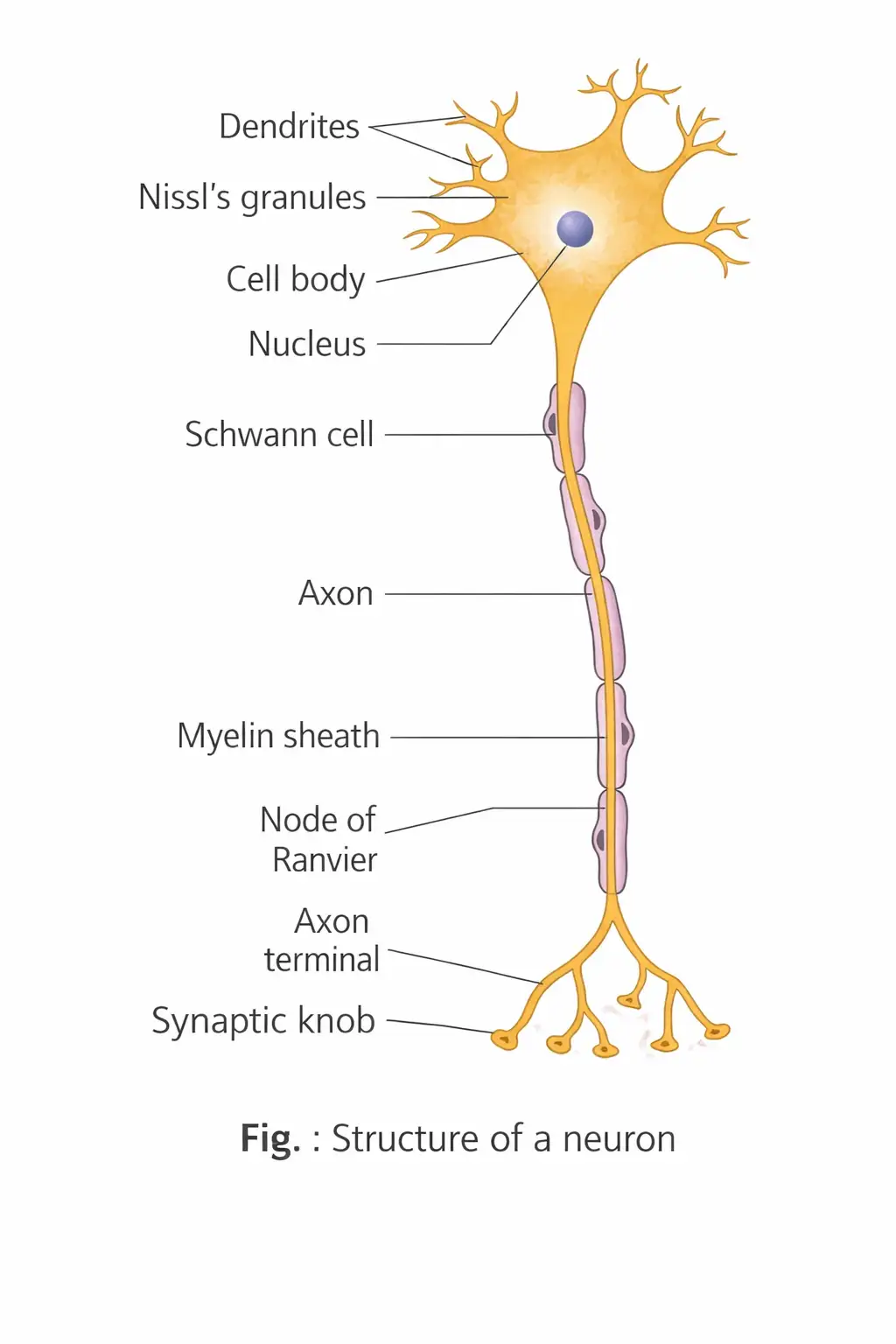
Neuron — A neuron is a microscopic, excitable cell and the structural and functional unit of the nervous system.
Main Parts
- Cell body: Contains cytoplasm, cell organelles and Nissl’s granules.
- Nissl’s granules: Irregular masses of rough endoplasmic reticulum with attached and free ribosomes and polysomes; they probably synthesise proteins.
- Dendrites: Short, repeatedly branched fibres containing Nissl’s granules; they conduct impulses towards the cell body.
- Axon: A long fibre with branched distal ends; it carries impulses away from the cell body to a synapse or neuromuscular junction.
- Synaptic knobs: Bulb-like endings of axon branches containing vesicles filled with neurotransmitters.
| Part | Main feature | Impulse direction |
|---|---|---|
| Dendrites | Short, branched; contain Nissl’s granules | Towards cell body |
| Axon | Long fibre; ends in branched terminals | Away from cell body |
Types of Neurons
| Type | Structure | Example / location |
|---|---|---|
| Unipolar | Cell body has one axon only | Usually found in the embryonic stage |
| Bipolar | One axon and one dendrite | Retina of the eye |
| Multipolar | One axon and two or more dendrites | Cerebral cortex |
| Pseudounipolar | One axon extends from the cell body and then divides into two | Dorsal root ganglion |
| Non-polar | Processes from the soma are equal; impulses can travel in any direction | Neurons of coelenterates |
| Type | Impulse direction | Nature / feature |
|---|---|---|
| Afferent (sensory) | Receptors to CNS | Dendrite terminals may be modified into receptors |
| Efferent (motor) | CNS to effectors such as muscles or glands | Axon terminals contact the motor end plate to form a neuromuscular junction |
Types of Axons
| Point | Myelinated | Unmyelinated |
|---|---|---|
| Schwann cells | Form a myelin sheath around the axon | Enclose the axon but do not form a myelin sheath |
| Nodes of Ranvier | Gaps between adjacent myelin sheaths | No myelin sheath gaps |
| Common location | Spinal and cranial nerves | Autonomic and somatic neural systems |
| Impulse conduction | Saltatory conduction; faster | Local currents pass along the axon; slower |
Generation and Conduction of a Nerve Impulse
Resting potential — The electrical potential difference across a resting neuronal membrane, with the inside negative relative to the outside.
Resting State
- Selective permeability: At rest, the axonal membrane is more permeable to \(K^+\) and nearly impermeable to \(Na^+\); it is impermeable to negatively charged proteins in the axoplasm.
- Ion distribution: Inside the axon, \(K^+\) and negatively charged proteins are concentrated, while \(Na^+\) is low. Outside, \(Na^+\) is high and \(K^+\) is low.
- Sodium–potassium pump: By active transport, it maintains ion gradients by moving \(3Na^+\) out for every \(2K^+\) moved into the cell.
- Polarisation: The outer membrane surface is positive and the inner surface negative; the resting potential inside is about \(-70\,\text{mV}\).
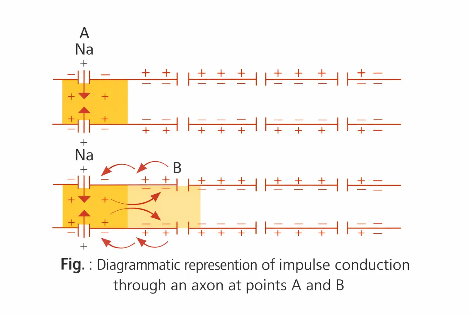
Action potential (nerve impulse) — The electrical potential difference at a stimulated site when depolarisation reverses the membrane’s polarity.
Action Potential and Propagation
- Stimulus: The stimulated membrane becomes highly permeable to \(Na^+\); rapid sodium influx reverses polarity.
- Depolarisation: The outside becomes negative and inside positive; the action-potential value is about \(+30\) to \(+45\,\text{mV}\).
- Local current: Current flows inside the axon from the excited site to the next site and returns along the outer surface, triggering depolarisation there.
- Propagation: This sequence repeats along the axon; nerve impulse conduction is unidirectional.
- Repolarisation: A brief increase in \(K^+\) permeability lets potassium diffuse out, restoring resting potential and responsiveness.
Refractory period — The period of complete inexcitability between depolarisation and repolarisation.
Saltatory conduction — In myelinated axons, the action potential appears to jump from one node of Ranvier to the next, making conduction faster than in non-myelinated axons.
Transmission of Impulses Across Synapses
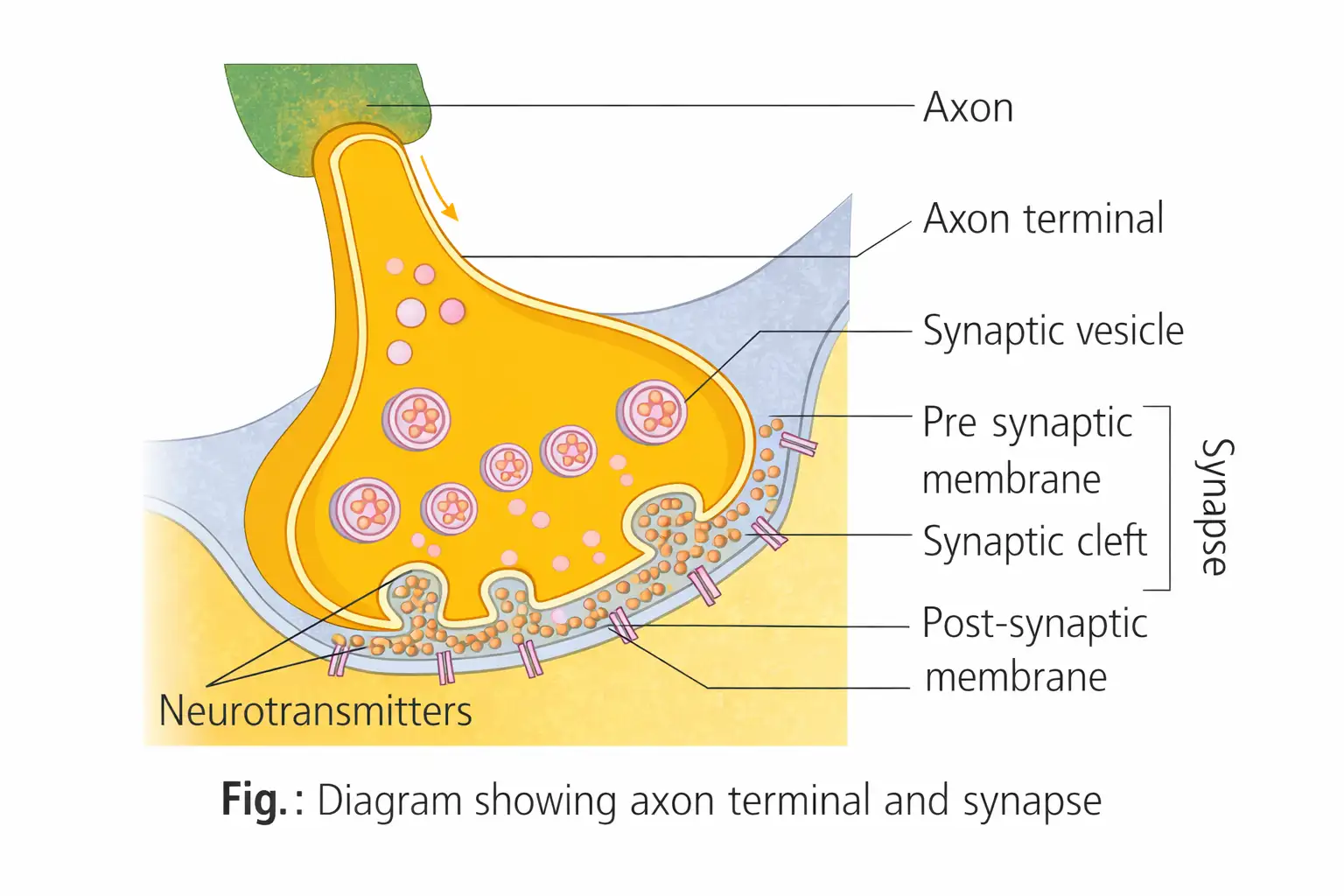
Synapse — A junction between presynaptic and postsynaptic neurons through which impulses pass; when present, the gap between them is the synaptic cleft.
Electrical Synapse
- Structure: Presynaptic and postsynaptic membranes lie very close together.
- Transmission: Electrical current passes directly from one neuron to the other, much like conduction along one axon.
- Occurrence and speed: Electrical synapses are rare in our nervous system and transmit faster than chemical synapses.
| Point | Electrical synapse | Chemical synapse |
|---|---|---|
| Structure | Pre- and postsynaptic membranes are very close | Membranes are separated by a fluid-filled synaptic cleft |
| Transmission | Electrical current flows directly between neurons | Neurotransmitters carry the signal across the cleft |
| Speed / occurrence | Faster; rare in our nervous system | Slower than electrical transmission |
Chemical Synapse Transmission
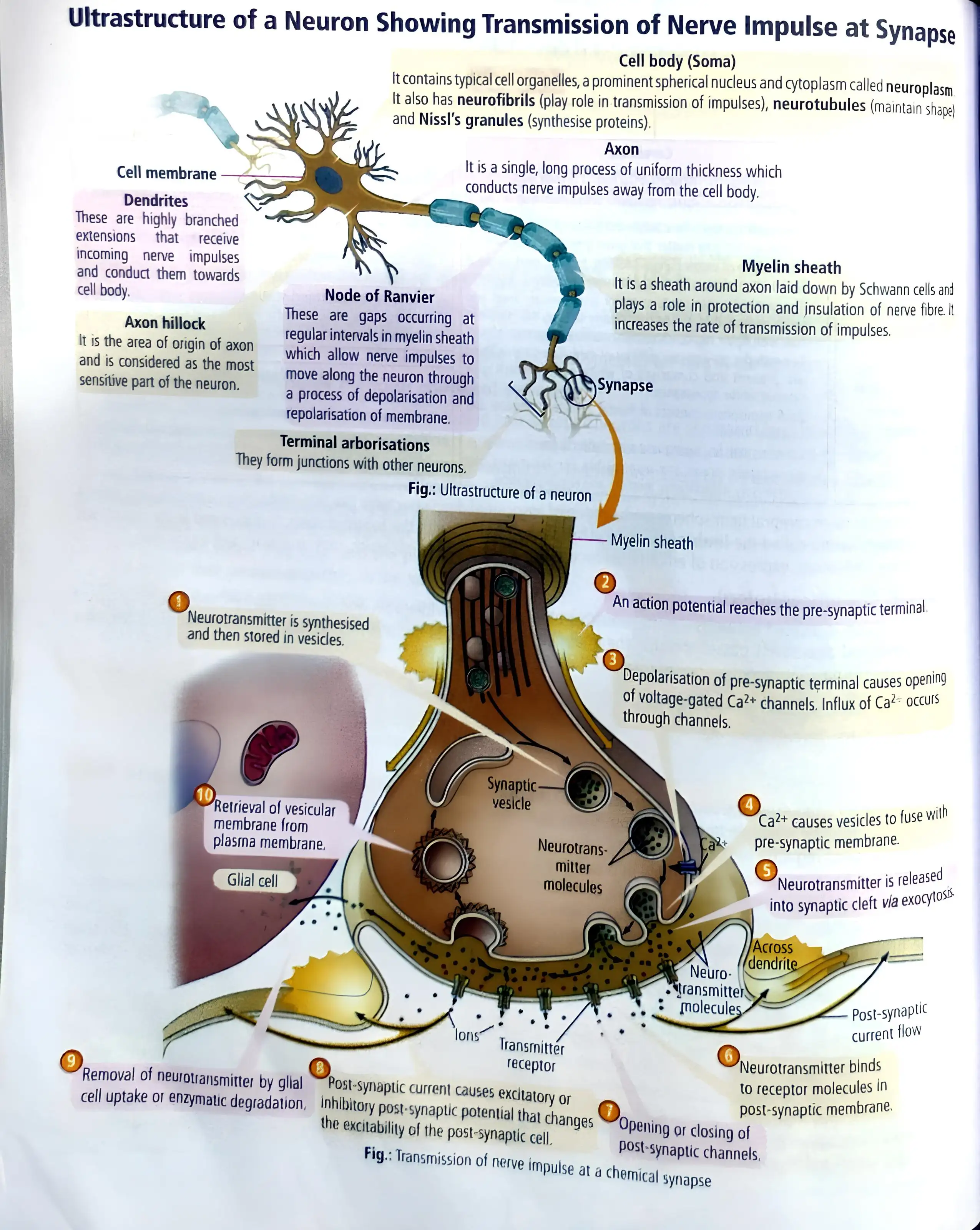
- Neurotransmitters: Synaptic vesicles in axon terminals store neurotransmitters. Acetylcholine is excitatory and GABA inhibitory; acetylcholine and noradrenaline are major neurotransmitters in vertebrates.
- Arrival: An action potential at the axon terminal stimulates vesicles to move towards and fuse with the plasma membrane.
- Release and binding: Neurotransmitters enter the synaptic cleft and bind to specific receptors on the postsynaptic membrane.
- Postsynaptic response: Receptor binding opens ion channels, allowing ions to enter and generate a new potential that may be excitatory or inhibitory.
How is the resting potential maintained in a neuron?
Outline impulse transmission across a chemical synapse.
Quick recap
- A neuron has a cell body, dendrites and an axon; dendrites conduct impulses towards the cell body and the axon away from it.
- Neurons are classified by their processes as unipolar, bipolar, multipolar, pseudounipolar or non-polar; functionally, they are afferent or efferent.
- At rest, the membrane is polarised; the inside is about \(-70\,\text{mV}\) and active transport by the pump moves \(3Na^+\) out for every \(2K^+\) in.
- Sodium influx depolarises a stimulated site; potassium efflux restores resting potential, and the impulse travels unidirectionally.
- Myelinated axons conduct faster by saltatory conduction between nodes of Ranvier.
- Electrical synapses transmit directly and faster; chemical synapses use neurotransmitters to produce an excitatory or inhibitory response.
Central Neural System
Brain: Role and Protection
Brain — The brain is the body’s central information-processing organ and command-and-control system.
- Controls: voluntary movements, body balance, vital involuntary organs such as the heart, lungs and kidneys, and human behaviour.
- Regulates: thermoregulation, hunger, thirst, 24-hour circadian rhythms and activities of several endocrine glands.
- Processes: vision, hearing, speech, memory, intelligence, emotions and thoughts.
- Protection: the skull encloses the brain; cranial meninges cover it inside the skull.
| Position | Layer |
|---|---|
| Outer | Dura mater |
| Middle; very thin | Arachnoid |
| Inner | Pia mater |
Cerebrospinal Fluid (CSF)
- Location: clear fluid in the brain’s ventricles and the subarachnoid space around the brain and spinal cord.
- Function: acts as a shock-absorbing medium for the brain.
- Major divisions: forebrain, midbrain and hindbrain.
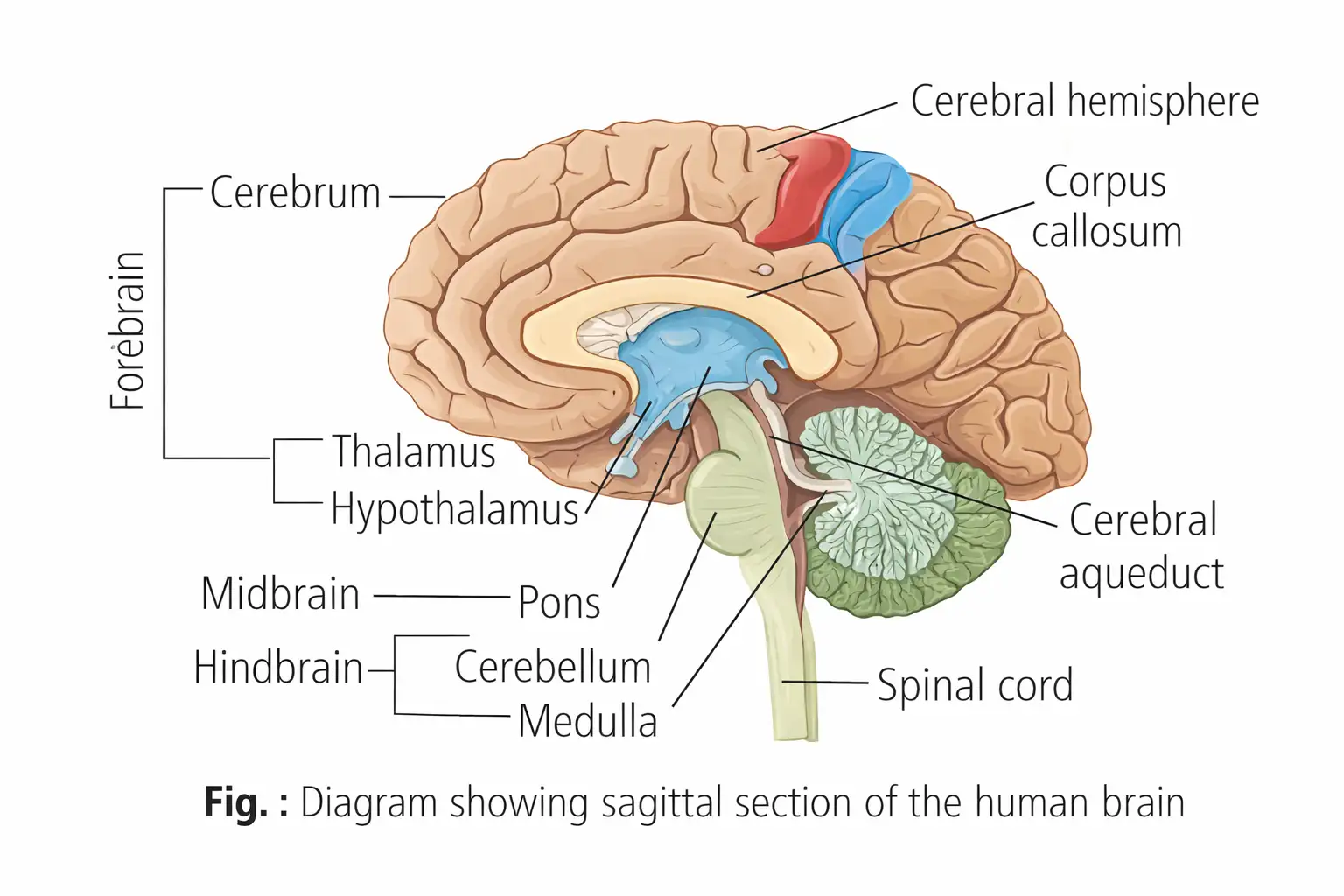
Forebrain (Prosencephalon)
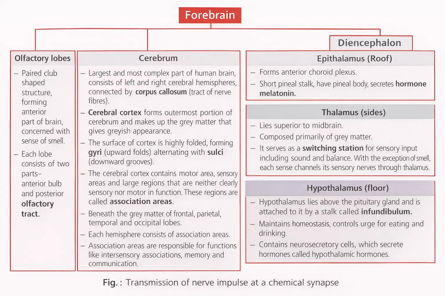
- Components: olfactory lobes, cerebrum and diencephalon.
- Limbic system: the inner part of the cerebral hemispheres and associated deep structures, including the amygdala and hippocampus, form the limbic lobe/system.
- Role: with the hypothalamus, it regulates sexual behaviour, emotional reactions such as excitement, pleasure, rage and fear, and motivation.
Midbrain (Mesencephalon)
- Location: between the thalamus/hypothalamus of the forebrain and the pons of the hindbrain.
- Cerebral aqueduct: a canal that passes through the midbrain.
- Corpora quadrigemina: four lobes on the dorsal midbrain; the superior colliculi are associated with sight and the inferior colliculi with hearing.
- Crura cerebri: relay impulses to and from the cerebrum, cerebellum, pons and medulla.
Hindbrain
- Components: pons, cerebellum and medulla oblongata (medulla).
- Pons: lies below the midbrain; its fibre tracts interconnect different brain regions. Its pneumotaxic centre regulates breathing.
- Cerebellum: its highly convoluted surface provides space for many neurons; it regulates and coordinates movement, posture and balance.
- Medulla: connected to the spinal cord; contains centres controlling respiration, cardiovascular reflexes and gastric secretions.
Brain Stem
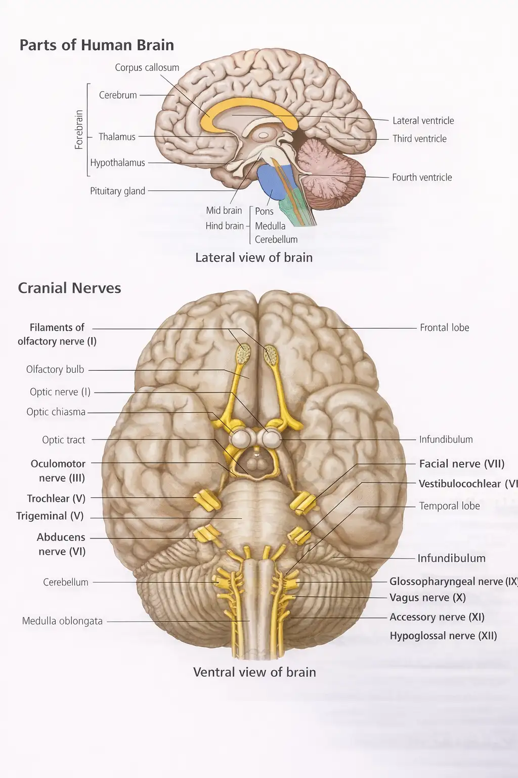
- Parts: midbrain, pons and medulla oblongata; it connects the brain to the spinal cord and supports life-sustaining functions.
- Functions: coordinates and integrates incoming information and forms the connection between the brain and spinal cord.
- Cranial nerves: serves as the entry or exit point for 10 of the 12 cranial nerves.
Spinal Cord
- Extent and size: extends from the medulla oblongata to the level of the second lumbar vertebra; it is about 42–45 cm long and weighs about 35 g.
- Functions: conducts impulses to and from the brain, controls most reflex activities and enables communication between spinal nerves and the brain.
- Tissue arrangement: internal grey matter is surrounded by outer white matter.
- Lower end: the spinal cord ends as the conus medullaris; a fine filament, the filum terminale, arises from it and anchors the cord within the vertebral column.
- Cauda equina: spinal nerves below the second lumbar vertebra are collectively called the cauda equina.
Quick recap
- The brain controls body functions and behaviour and processes sensory, cognitive and emotional information.
- The brain is protected by the skull, three cranial meninges and shock-absorbing CSF in the ventricles and subarachnoid space.
- The brain has three divisions: forebrain, midbrain and hindbrain; the forebrain includes olfactory lobes, cerebrum and diencephalon.
- The limbic system with the hypothalamus helps regulate emotions, sexual behaviour and motivation.
- The pons, cerebellum and medulla are linked to breathing, movement, posture, balance and vital involuntary functions.
- The brain stem comprises the midbrain, pons and medulla; it integrates incoming information and is the entry or exit point for 10 of 12 cranial nerves.
- The spinal cord extends from the medulla to the second lumbar vertebra, conducts impulses and reflexes, and has internal grey matter with outer white matter.
NEET Zoology — ALL CHAPTERS
1
Structural Organisation in Animals
2
Breathing and Exchange of Gases
3
Body Fluids and Circulation
4
Excretory Products and their Elimination
5
Locomotion and Movement
6
Neural Control and Coordination
7
Human Reproduction
8
Reproductive Health
9
Evolution
10
Microbes in Human Welfare
11
Organism and Population
12
Ecosystem
13
Biodiversity and Conservation