NEET Ug . NEET Zoology . Zoology . Excretory Products and Their Elimination
Chapter 4: Excretory Products and their Elimination
Ch 4
NEET Zoology
NEET Ug
Urine Formation
Excretion and Nitrogenous Wastes
| Feature | Ammonotelic | Ureotelic | Uricotelic |
|---|---|---|---|
| Main waste | Ammonia | Urea | Uric acid |
| Water requirement | Very large | Less than for ammonia | Least |
| Excretion | Diffuses across body surface or fish gills, often as ammonium ion | Ammonia is converted to urea in the liver; urea enters the blood, is filtered by the kidneys and excreted | Excreted as a pellet or paste |
| Toxicity | Most toxic | Less toxic than ammonia | Least toxic |
| Examples | Many bony fishes, aquatic insects and aquatic amphibians | Mammals, marine fishes and terrestrial amphibians | Reptiles, birds, insects and land snails |
| Excretory organ | Animal |
|---|---|
| Contractile vacuoles and plasma membrane | Protozoans |
| General body surface | Hydra and sponges |
| Renette cells | Aschelminthes |
| Flame cells (solenocytes and protonephridia) | Planarians (Platyhelminthes) |
| Nephridia and chloragogen cells | Earthworm (Annelida) |
| Malpighian tubules | Insects (Arthropoda) |
| Coxal glands | Spiders (Arachnida) |
| Antennal glands or green glands | Prawns (Crustacea) |
| Keber’s organ and organs of Bojanus | Molluscs |
| Kidneys | Vertebrates |
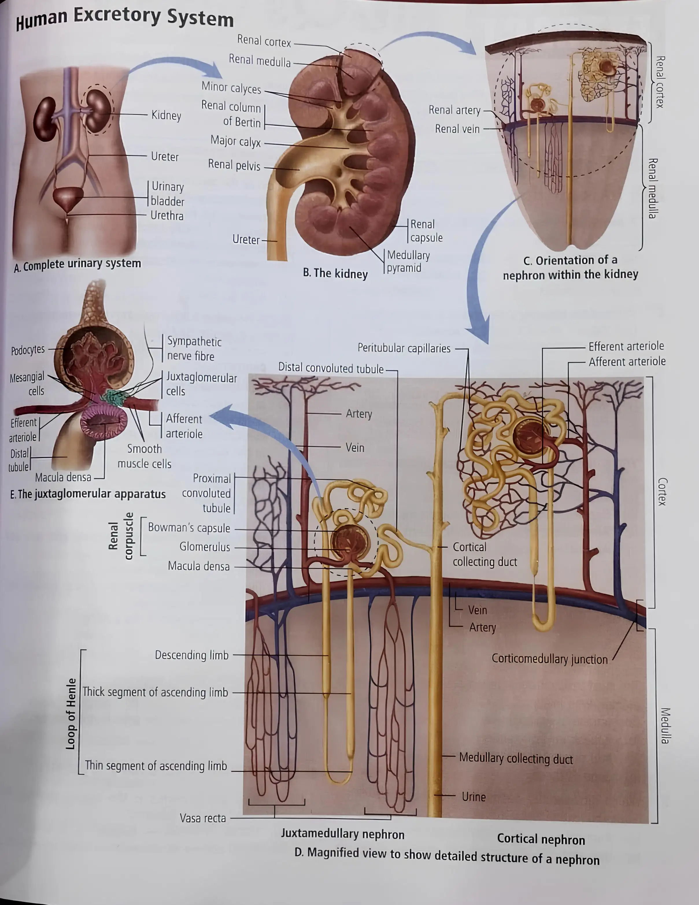
Human Excretory System
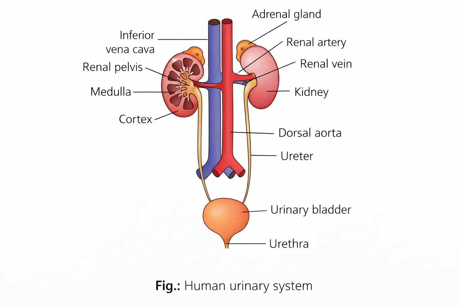
Kidney Structure
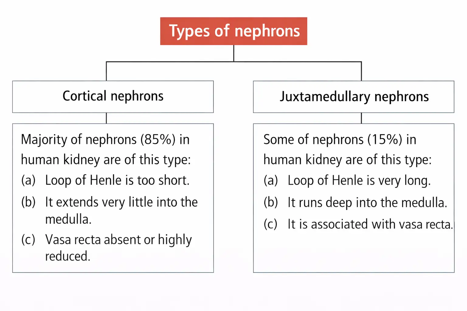
Nephron: Structure and Types
| Type | Key feature or occurrence |
|---|---|
| Archinephros (archinephric) | Basic ancestral form; found in larvae of some cyclostomes, such as Myxine |
| Pronephros (pronephric) | Most primitive excretory organ; develops embryonically in some vertebrates and represents the first stage of kidney development |
| Mesonephros (mesonephric) | Middle kidney; main excretory organ of aquatic vertebrates and a temporary kidney in higher vertebrates |
| Metanephros (metanephric) | Most advanced kidney, with a loop of Henle; functional in reptiles, birds and mammals |
Nephron Regions and Associated Blood Vessels
Stages of Urine Formation
Glomerular Filtration or Ultrafiltration
Glomerular filtration rate (GFR) — The volume of filtrate formed by the kidneys per unit time.
Normal Filtration Values
- GFR: About 125 mL/min, or approximately 180 L/day, in a healthy individual.
- Renal plasma flow (RPF): About 650–700 mL of plasma passes through the kidneys per minute.
Regulation of GFR and Effective Filtration Pressure
| Pressure | Effect | Value |
|---|---|---|
| Glomerular blood hydrostatic pressure (GBHP) | Promotes filtration; chief determinant of EFP, partly due to the narrower efferent arteriole | 60 mmHg |
| Blood colloidal osmotic pressure (BCOP) | Opposes filtration because of plasma proteins | 32 mmHg |
| Capsular hydrostatic pressure (CHP) | Opposes filtration because filtrate in Bowman’s capsule exerts pressure | 18 mmHg |
Net pressure promoting filtration; the normal value is 10 mmHg.
Tubular Reabsorption
Tubular Secretion
Name the three processes involved in urine formation.
What is the normal GFR, and how is it regulated when it falls?
Calculate EFP using the given pressure values.
Quick recap
- Excretion removes metabolic wastes; ammonia, urea and uric acid are the principal nitrogenous wastes.
- The nephron is the kidney’s structural and functional unit, with a renal corpuscle and renal tubule.
- Urine formation involves filtration, reabsorption and secretion.
- The filtration barrier retains most plasma proteins; normal GFR is about 125 mL/min.
- JGA cells release renin when GFR falls, helping restore filtration.
- About 99% of filtrate materials are reabsorbed; secretion supports ionic and acid–base balance.
Function of the Tubules
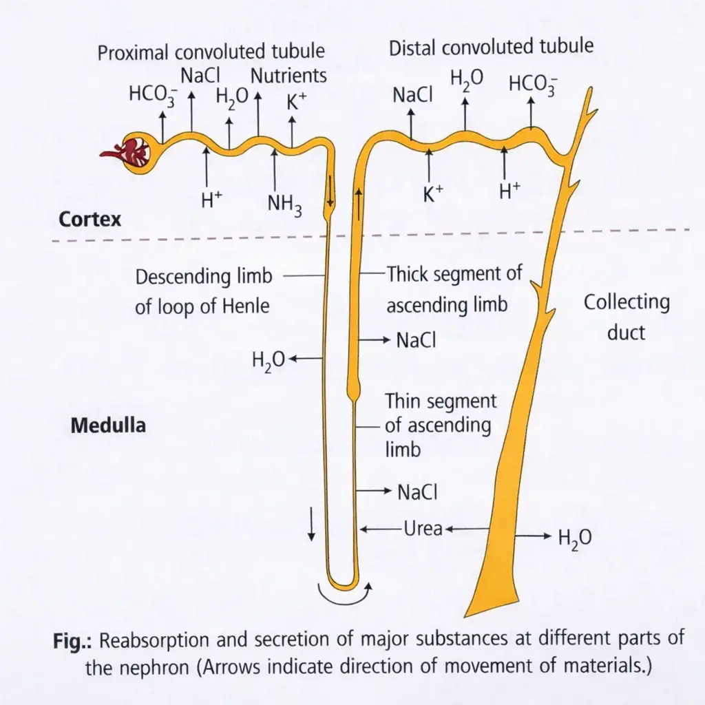
Proximal Convoluted Tubule (PCT)
- Lining: Simple cuboidal brush-border epithelium increases the surface area available for reabsorption.
- Reabsorption: Nearly all essential nutrients and about 70–80% of filtered water and electrolytes are reabsorbed here; reabsorption is maximal in the PCT.
- Transport: Glucose and amino acids are reabsorbed by active transport; water is reabsorbed by osmosis.
- Secretion and balance: Selective secretion of \(H^+\), ammonia and \(K^+\), with absorption of \(HCO_3^-\), helps maintain body-fluid pH and ionic balance.
Henle’s Loop
| Feature | Descending limb | Ascending limb |
|---|---|---|
| Water permeability | Permeable to water | Impermeable to water |
| Electrolyte movement | Almost impermeable to electrolytes | Electrolytes move out actively or passively |
| Effect on filtrate | Water leaves, concentrating the filtrate as it descends | Electrolytes leave, diluting the concentrated filtrate as it ascends |
- Ascending-limb reabsorption: Reabsorption is minimal here, but electrolyte transport helps maintain the high osmolarity of the medullary interstitial fluid.
Distal Convoluted Tubule (DCT)
- Conditional reabsorption: Reabsorption of \(Na^+\) and water occurs according to the body’s needs.
- Other reabsorption: \(HCO_3^-\) is also reabsorbed.
- Secretion and balance: Selective secretion of \(H^+\), \(K^+\) and \(NH_3\) helps maintain blood pH and sodium–potassium balance.
Collecting Duct
- Path: Extends from the kidney cortex into the inner medulla.
- Water reabsorption: Large amounts of water may be reabsorbed, producing concentrated urine.
- Medullary osmolarity: A small amount of urea passes into the medullary interstitium, helping maintain its osmolarity.
- Secretion and balance: Selective secretion of \(H^+\) and \(K^+\) contributes to blood pH and ionic balance.
Quick recap
- PCT has brush-border cells and carries out the nephron’s greatest reabsorption.
- The PCT reabsorbs nearly all essential nutrients and about 70–80% of water and electrolytes.
- The Henle’s loop descending limb loses water; its ascending limb loses electrolytes and is impermeable to water.
- DCT conditionally reabsorbs \(Na^+\) and water and selectively secretes ions to help maintain blood balance.
- The collecting duct can reabsorb substantial water and contributes to concentrated urine and medullary osmolarity.
Mechanism of Concentration of the Filtrate
Counter-current arrangement
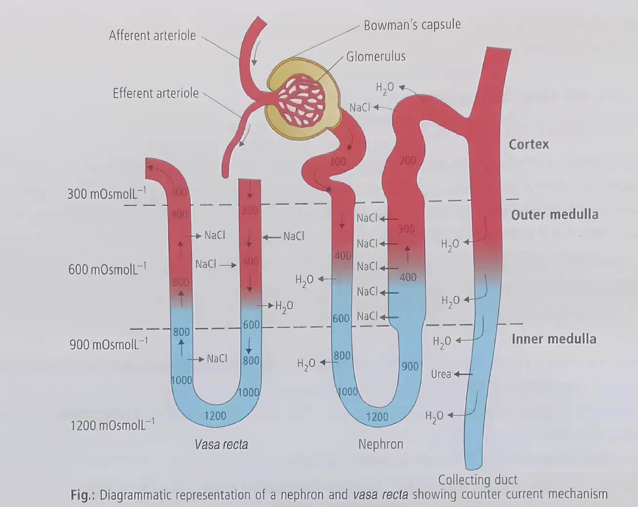
Counter-current mechanism — The special transport and exchange of substances between the loop of Henle and vasa recta, which have flows in opposite directions, helps maintain the medullary osmotic gradient.
Where it occurs and how the flows oppose
- Location: The concentration mechanism operates in the kidney medulla, involving the loop of Henle and vasa recta.
- Loop of Henle: Filtrate flows in opposite directions through its descending and ascending limbs.
- Vasa recta: Blood flows in opposite directions through its two limbs.
- Proximity: The close arrangement of the loop of Henle and vasa recta supports the medullary osmotic gradient.
Medullary osmotic gradient
Gradient and solute exchange
- Direction: Osmolarity increases from about \(300\ \text{mOsmol L}^{-1}\) in the cortex to about \(1200\ \text{mOsmol L}^{-1}\) in the inner medulla.
- Main solutes: Sodium chloride (\(\ce{NaCl}\)) and urea mainly create this gradient.
- NaCl exchange: NaCl moves from the ascending limb of Henle’s loop to the descending vasa recta, then returns to the medullary interstitium from the ascending vasa recta.
- Urea movement: A small amount of urea enters the thin ascending limb of Henle’s loop and is transported back to the interstitium by the collecting tubule.
How the gradient concentrates urine
- Opposite flows and solute exchange help maintain high osmolarity in the medullary interstitium.
- The medullary gradient promotes water movement out of the collecting tubule.
- Water reabsorption concentrates the filtrate, producing concentrated urine.
Urine concentration and loop length
- Human kidneys: They can produce urine nearly four times as concentrated as the initial filtrate.
- Loop length: Across mammals, urine-concentrating ability is directly related to the length of the loop of Henle.
- Water conservation: A longer loop allows more water reabsorption, producing more concentrated urine.
What is the counter-current mechanism, and how does it help concentrate urine?
How is the length of the loop of Henle related to urine concentration?
Quick recap
- The concentration mechanism occurs in the kidney medulla and involves the loop of Henle and vasa recta.
- Filtrate and blood flow in opposite directions in the two structures, supporting counter-current exchange.
- Medullary osmolarity rises from about \(300\ \text{mOsmol L}^{-1}\) in the cortex to about \(1200\ \text{mOsmol L}^{-1}\) in the inner medulla.
- \(\ce{NaCl}\) and urea are the main solutes contributing to the gradient.
- The gradient promotes water reabsorption from the collecting tubule and concentrates urine.
- A longer loop of Henle is associated with more concentrated urine.
Regulation of Kidney Function
Hormonal Feedback Regulation
Antidiuretic Hormone (ADH)
Diuretics — Drugs that increase the production of dilute urine, helping prevent excessive water retention and tissue swelling (oedema).
| Substance | Effect |
|---|---|
| Alcohol | Inhibits ADH release, increasing water loss in urine. |
| Caffeine | Interferes with ADH action and sodium reabsorption, increasing water loss in urine. |
Diabetes insipidus — A disorder caused by ADH deficiency; urine output may reach 20–25 litres per day.
Juxtaglomerular Apparatus (JGA) and Renin–Angiotensin Mechanism
Renin–Angiotensin Mechanism
- Decreased glomerular blood flow, glomerular blood pressure or GFR activates JG cells in the JGA.
- Activated JG cells release renin.
- Renin converts blood angiotensinogen into angiotensin I, which is then converted into angiotensin II.
- Angiotensin II, a powerful vasoconstrictor, raises glomerular blood pressure and GFR; it also stimulates the adrenal cortex to release aldosterone.
- Aldosterone promotes sodium (\(Na^+\)) and water reabsorption in the distal parts of the tubule, increasing blood pressure and GFR.
Atrial Natriuretic Factor (ANF)
Quick recap
- Osmoreceptors detect changes in blood and body-fluid volume and ionic concentration; fluid loss triggers ADH release from the neurohypophysis.
- ADH increases water reabsorption in distal tubules and constricts blood vessels, potentially increasing GFR.
- Diuretics increase dilute urine production; alcohol and caffeine increase urinary water loss through different effects.
- ADH deficiency causes diabetes insipidus, with urine output that may reach 20–25 litres per day.
- Reduced glomerular blood flow, pressure or GFR triggers renin release and the renin–angiotensin mechanism.
- Angiotensin II raises glomerular pressure and stimulates aldosterone; aldosterone promotes distal-tubule sodium and water reabsorption.
- Increased atrial blood flow releases ANF, which causes vasodilation and inhibits renin release.
Micturition
Micturition and the Micturition Reflex
Micturition Reflex: Sequence
- Bladder fills with urine → bladder wall stretches
- Stretch receptors signal the CNS
- CNS sends motor messages → bladder smooth muscle contracts and urethral sphincter relaxes
- Urine is expelled
Urine: Characteristics and Composition
Urine Analysis and Clinical Significance
Describe the micturition reflex.
Quick recap
- Micturition is the expulsion of urine; its neural mechanism is the micturition reflex.
- Bladder stretch signals the CNS, which contracts bladder muscle and relaxes the urethral sphincter.
- An adult excretes about 1–1.5 L of urine and 25–30 g of urea daily.
- Urine is slightly acidic (average pH 6.0); urochrome gives it its yellow colour.
- Bacteria convert urea to ammonia in standing urine, causing a strong smell.
- Glycosuria and ketonuria may indicate diabetes mellitus; albuminuria and pyuria describe other urine abnormalities.
Role of Other Organs in Excretion
Lungs and Liver
Skin Glands
Saliva
Quick recap
- Lungs eliminate CO₂ at about 200 mL/min and remove water.
- The liver secretes several wastes and other substances in bile; most leave with digestive wastes.
- Sweat contains NaCl, small amounts of urea and lactic acid; its main function is cooling.
- Sebum carries sterols, hydrocarbons and waxes and protects the skin with an oily covering.
- Saliva can remove small amounts of nitrogenous wastes.
Disorders of the Excretory System
Uremia and Hemodialysis
Kidney Transplantation
Renal Calculi
Glomerulonephritis
Quick recap
- Uremia is the harmful accumulation of urea in blood due to kidney malfunction.
- Hemodialysis removes blood wastes across a porous cellophane membrane into waste-free dialysing fluid.
- Heparin prevents clotting during dialysis; anti-heparin is added before blood returns to the body.
- Kidney transplantation uses a compatible donor kidney; a close relative is preferred to reduce rejection risk.
- Renal calculi are crystallised salt masses in the kidney; causes include dehydration and metabolic or hormonal disturbances.
- Glomerulonephritis is inflammation of the kidney glomeruli.
NEET Zoology — ALL CHAPTERS
1
Structural Organisation in Animals
2
Breathing and Exchange of Gases
3
Body Fluids and Circulation
4
Excretory Products and their Elimination
5
Locomotion and Movement
6
Neural Control and Coordination
7
Human Reproduction
8
Reproductive Health
9
Evolution
10
Microbes in Human Welfare
11
Organism and Population
12
Ecosystem
13
Biodiversity and Conservation