NEET Ug . NEET Zoology . Zoology . Breathing and Exchange of Gases
Chapter 2: Breathing and Exchange of Gases
Ch 2
NEET Zoology
NEET Ug
Respiratory Organs
Introduction: Breathing and Respiration
Why organisms need gas exchange
- Oxygen use: Cells use oxygen to oxidise food molecules such as glucose, fatty acids and amino acids, releasing energy for life activities.
- Carbon dioxide: Catabolic reactions produce carbon dioxide, which must be removed from the body.
- Reductionist approach: Studying life through its physico-chemical components is called the reductionist approach; neither this approach alone nor an organism-level approach alone fully explains living phenomena.
Breathing — The intake of fresh air and removal of carbon-dioxide-rich air from the body.
Respiration — The exchange of environmental oxygen with the carbon dioxide produced in cells during food oxidation.
| Point | Breathing | Respiration |
|---|---|---|
| What it involves | Air enters and leaves the body. | Gas exchange, gas transport and cellular use of oxygen, with carbon dioxide release. |
| Where it occurs | Respiratory passages and lungs. | Across respiratory surfaces, in blood and tissues, and in cells. |
Respiratory Organs in Animals
- Variation: Respiratory mechanisms differ among animal groups, mainly according to habitat and level of organisation.
| Respiratory organ / mode | Examples |
|---|---|
| General body surface — simple diffusion | Sponges, coelenterates, flatworms |
| Moist skin — cutaneous respiration | Earthworms, frogs |
| Tracheal tubes — tracheal respiration | Insects |
| Gills — branchial respiration | Aquatic arthropods, molluscs, fishes |
| Lungs — pulmonary respiration | Amphibians, reptiles, birds, mammals |
Human Respiratory System
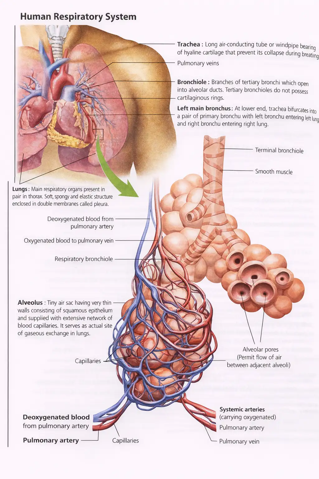
Air passage and branching
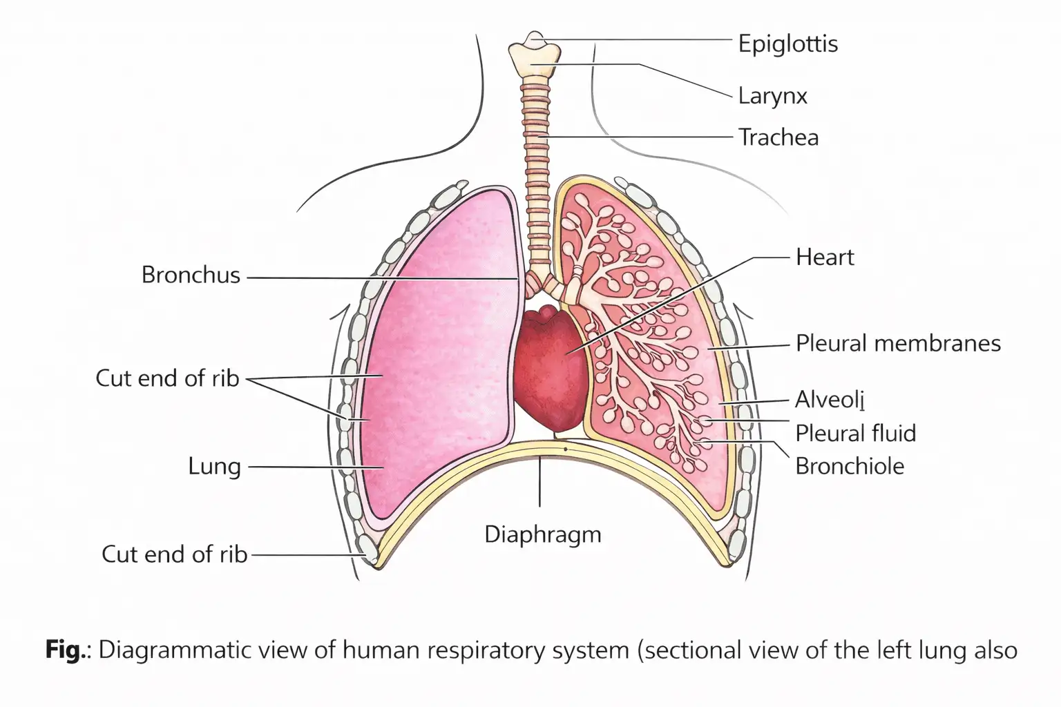
- External nostrils → nasal passage → nasal chamber → pharynx.
- Pharynx → glottis (slit-like opening) → larynx → trachea.
- Trachea divides into right and left primary bronchi at the fifth thoracic vertebra.
- Primary bronchi → secondary and tertiary bronchi → bronchioles → terminal bronchioles → alveoli.
Airway structures and alveoli
- Pharynx: A shared passage for food and air.
- Epiglottis: A thin, elastic, cartilaginous flap that covers the glottis during swallowing, preventing food from entering the larynx.
- Larynx: A cartilaginous box that produces sound; also called the sound box.
- Cartilage support: Incomplete cartilaginous rings support the trachea, bronchi up to the tertiary level, and the initial bronchioles.
- Alveoli: Very thin-walled, irregular, vascularised air sacs arising from terminal bronchioles; close contact with capillaries enables gas exchange.
- Number: Human lungs contain approximately 300 million alveoli.
Lungs, Pleura and Respiratory Parts
Pleural membranes
- Pleura: Each lung is enclosed by a double-layered membrane; the outer layer contacts the thoracic lining and the inner layer contacts the lung surface.
- Pleural cavity: The narrow space between the two pleural layers contains pleural fluid.
- Pleural fluid: Lubricates the layers so they slide over one another with little friction as the lungs expand and contract.
| Point | Conducting part | Respiratory part |
|---|---|---|
| Extent | External nostrils to terminal bronchioles. | Alveoli and their ducts. |
| Main role | Carries air to alveoli; filters foreign particles, humidifies air and brings it to body temperature. | Site of actual diffusion of oxygen and carbon dioxide between air and blood. |
Alveolar Cells and Surfactant
| Cell type | Main feature |
|---|---|
| Type I pneumocytes | Squamous cells; mainly responsible for gaseous exchange. |
| Type II pneumocytes | Larger and fewer; secrete lecithin, a phospholipid that acts as surfactant. |
- Surfactant: A lipid that lowers surface tension and prevents alveoli from collapsing during respiration.
- Development: Surfactant secretion develops only by the eighth or ninth month of pregnancy.
- Clinical significance: In some newborns, an absent lecithin film can cause lung collapse and may be fatal.
Thoracic Chamber and Lung Movement
- Thoracic chamber: An airtight chamber containing the lungs; bounded dorsally by the vertebral column, ventrally by the sternum, laterally by the ribs and below by the dome-shaped diaphragm.
- Volume relationship: The lungs are arranged so that a change in thoracic-cavity volume is reflected in the pulmonary cavity and lungs.
Steps in Respiration
Overall sequence
- Pulmonary ventilation moves air between the atmosphere and alveoli.
- Oxygen and carbon dioxide diffuse across the alveolar membrane.
- Blood transports the gases.
- Oxygen and carbon dioxide diffuse between blood and tissues.
- Cells use oxygen in catabolic reactions and release carbon dioxide (cellular respiration).
Differentiate the conducting part from the respiratory part of the human respiratory system.
What is the role of surfactant, and why is its deficiency dangerous in newborns?
Quick recap
- Breathing is air intake and removal; respiration also includes gas exchange, transport and cellular oxygen use.
- Respiratory organs vary with animal group and include body surface, moist skin, tracheal tubes, gills and lungs.
- Air travels from the nostrils through the conducting passages to alveoli, the main site of gas exchange.
- Type I pneumocytes exchange gases; Type II pneumocytes secrete lecithin-based surfactant that helps prevent alveolar collapse.
- Pleural fluid lubricates the double-layered pleura, while thoracic-cavity volume changes are reflected in the lungs.
- Respiration proceeds through ventilation, alveolar diffusion, blood transport, tissue diffusion and cellular use of oxygen.
Mechanism of Breathing
Basic Principle
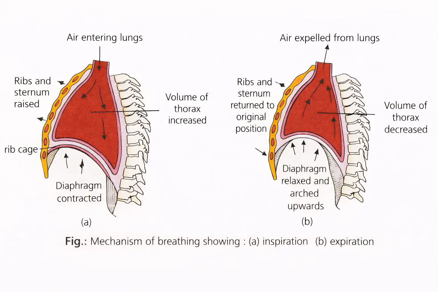
- Breathing has two stages: inspiration draws atmospheric air in; expiration releases alveolar air out.
- Pressure gradient: Air moves into or out of the lungs because pressure differs between the lungs and atmosphere.
- Main muscles: The diaphragm and the external and internal intercostal muscles help create this pressure gradient.
Inspiration
Inspiration — Air enters the lungs when intrapulmonary pressure becomes lower than atmospheric pressure.
How Inspiration Occurs
- Diaphragm contracts, flattens and lowers → thoracic volume increases along the anteroposterior axis.
- External intercostal muscles contract → ribs and sternum lift → thoracic volume increases along the dorsoventral axis.
- Thoracic volume increases → pulmonary volume increases → intrapulmonary pressure falls below atmospheric pressure.
- Air flows from outside into the lungs; abdominal muscles relax, allowing the diaphragm to compress abdominal organs. They play a passive role in inspiration.
Air Pathway During Inspiration
- External nares → nasal cavities → internal nares → pharynx → glottis → larynx → trachea → bronchi → bronchioles → alveolar ducts → alveoli.
Expiration
Expiration — Air leaves the lungs when intrapulmonary pressure rises above atmospheric pressure.
How Expiration Occurs
- Diaphragm and intercostal muscles relax → diaphragm and sternum return to normal positions.
- Thoracic volume decreases → pulmonary volume decreases → intrapulmonary pressure rises slightly above atmospheric pressure.
- Air is expelled from the lungs.
Air Pathway During Expiration
- Alveoli → alveolar ducts → bronchioles → bronchi → trachea → larynx → glottis → pharynx → internal nares → nasal cavities → external nares → outside.
- Forceful breathing: Additional abdominal muscles can increase the strength of inspiration and expiration.
| Point | Inspiration | Expiration |
|---|---|---|
| Muscle action | Active: involves muscle contraction. | Usually passive: results from muscle relaxation. |
| Intrapulmonary pressure | Below atmospheric pressure | Slightly above atmospheric pressure |
| Thoracic and pulmonary volume | Increase | Decrease |
| Air movement | Into the lungs | Out of the lungs |
Breathing Rate and Timing
- Average rate: A healthy human breathes about 12–16 times per minute.
- Duration: Inspiration takes about 2 seconds; expiration takes about 3 seconds.
Respiratory Volumes and Capacities
Respiratory volumes — Quantities of air that the lungs can inhale, hold or expel under different conditions.
Pulmonary capacities — Combinations formed by adding respiratory volumes; they are useful in clinical diagnosis.
Measurement and Variation
- Sex differences: Respiratory volumes and capacities are about 20%–25% lower in women than in men.
- Physical build: Values are generally greater in athletic people than in asthenic (thin, non-exercising) people.
- Spirometer: All volumes and capacities can be measured with a simple spirometer except residual volume (RV) and functional residual capacity (FRC).
| Type | Approximate quantity | Definition or formula |
|---|---|---|
| Tidal volume (TV) | 500 mL | Air inspired and expired during normal, effortless breathing. |
| Inspiratory reserve volume (IRV) | 2,500–3,000 mL | Extra air that can be forcibly inspired after a normal inspiration. |
| Expiratory reserve volume (ERV) | 1,000–1,100 mL | Extra air that can be expelled after a forcible expiration. |
| Residual volume (RV) | 1,100–1,200 mL | Air remaining in the lungs after a forceful expiration. |
| Vital capacity (VC) | 4,000–4,600 mL | Maximum volume of air inspired and expired; \(VC = TV + IRV + ERV\). |
| Inspiratory capacity (IC) | 3,000–3,500 mL | Air that can be inhaled after a normal expiration; \(IC = TV + IRV\). |
| Expiratory capacity (EC) | 1,500–1,600 mL | Air that can be expired after a normal inspiration; \(EC = TV + ERV\). |
| Functional residual capacity (FRC) | 2,100–2,300 mL | Air remaining in the lungs after a normal expiration; \(FRC = ERV + RV\). |
| Total lung capacity (TLC) | 5,100–5,800 mL | Total air in the lungs and respiratory passage after maximum inspiration; \(TLC = VC + RV\). |
Remember that RV remains after forceful expiration, while FRC remains after normal expiration; neither is measured directly by a simple spirometer.
Quick recap
- Breathing consists of inspiration and expiration, driven by pressure differences between the lungs and atmosphere.
- Inspiration is active: muscle contraction increases thoracic and pulmonary volumes and lowers intrapulmonary pressure.
- Abdominal muscles relax and play a passive role during inspiration; additional abdominal muscles can strengthen forceful breathing.
- Expiration is usually passive: muscle relaxation reduces lung volume and raises intrapulmonary pressure.
- A healthy person breathes about 12–16 times per minute; inspiration lasts about 2 seconds and expiration about 3 seconds.
- Pulmonary capacities combine respiratory volumes; for example, \(VC = TV + IRV + ERV\).
- A simple spirometer cannot measure RV or FRC.
Exchange of Gases
Sites and Direction of Gas Exchange
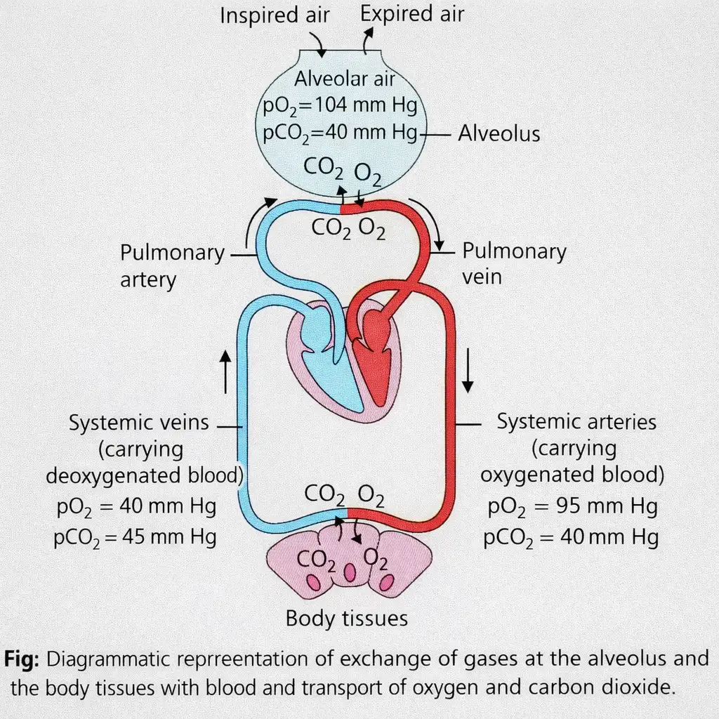
- Main site: Alveoli are the primary sites of gas exchange.
- Other site: Gases are also exchanged between blood and body tissues.
- Oxygen pathway: Oxygen diffuses from alveoli into blood, then from blood to tissues.
- Carbon dioxide pathway: Carbon dioxide diffuses from tissues into blood, then from blood to alveoli.
- Mechanism: Both gases move by simple diffusion, mainly down their partial-pressure or concentration gradients.
Partial Pressure and Diffusion Gradients
Partial pressure — The pressure contributed by an individual gas in a mixture; oxygen and carbon dioxide partial pressures are written as \(pO_2\) and \(pCO_2\), respectively.
| Gas | Atmosphere | Alveoli | Deoxygenated blood | Oxygenated blood | Tissues |
|---|---|---|---|---|---|
| \(O_2\) | 159 | 104 | 40 | 95 | 40 |
| \(CO_2\) | 0.3 | 40 | 45 | 40 | 45 |
- Oxygen gradient: Higher \(pO_2\) in alveoli drives oxygen into deoxygenated blood; blood then carries it to tissues with lower \(pO_2\).
- Carbon dioxide gradient: Higher \(pCO_2\) in tissues drives carbon dioxide into blood and onward to alveoli.
- Diffusion rate: Gas solubility and the thickness of the diffusion membrane affect how rapidly a gas diffuses.
- Carbon dioxide: Its solubility is about 20–25 times that of oxygen, so more \(CO_2\) diffuses per unit difference in partial pressure.
Diffusion Membrane and Blood Gas Levels
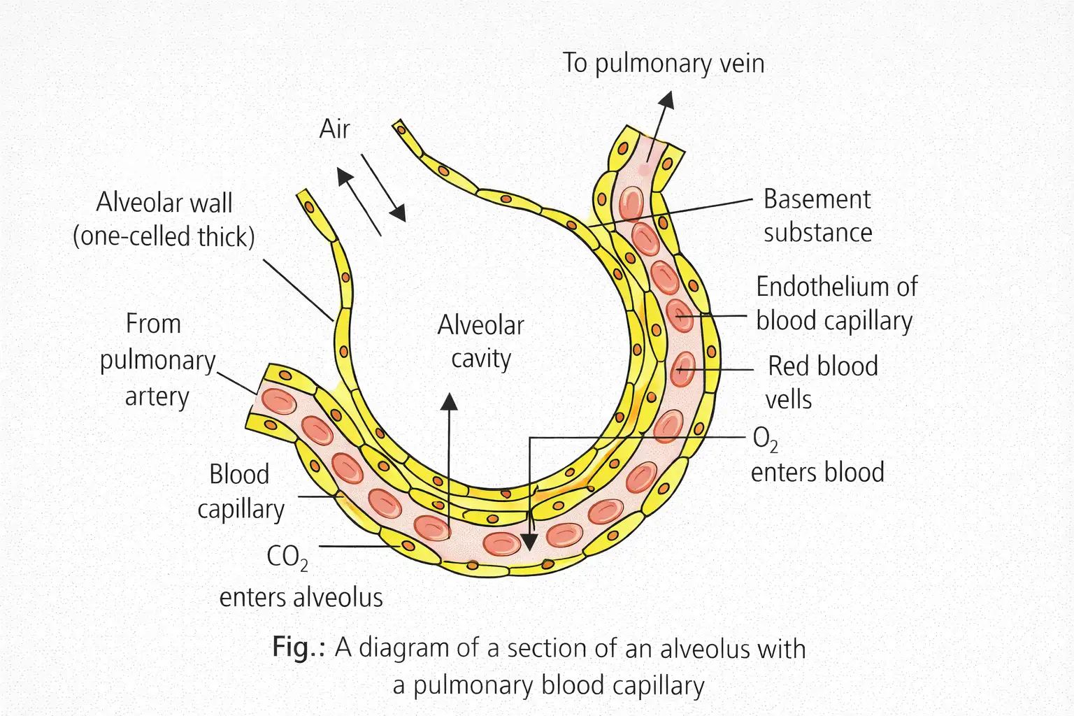
- Layers: The diffusion membrane consists of thin squamous alveolar epithelium, alveolar-capillary endothelium, and basement substance between them.
- Basement substance: It includes the thin basement membranes supporting the alveolar epithelium and surrounding the capillary endothelial cells.
- Thickness: The complete membrane is much less than \(1\) mm thick, providing a short diffusion path.
- Blood leaving alveoli: Arterial blood has almost the same gas partial pressures as alveolar air: \(pO_2 \approx 104\) mm Hg and \(pCO_2 \approx 40\) mm Hg.
Requirements for Efficient Gas Exchange
- Large surface area: Provides extensive area for gas exchange.
- Short diffusion path: Keeps the distance between alveolar air and blood very small.
- Partial-pressure gradients: Drive oxygen from alveoli to blood and carbon dioxide from blood to alveoli.
- Respiratory pigment: Haemoglobin increases the blood's carrying capacity for oxygen and carbon dioxide.
- Respiratory membrane: Must be thin, moist and large.
Quick recap
- Alveoli are the main gas-exchange sites; exchange also occurs between blood and tissues.
- Gases diffuse down their partial-pressure gradients: oxygen toward tissues and carbon dioxide toward alveoli.
- Diffusion depends on gas solubility and membrane thickness; carbon dioxide is 20–25 times more soluble than oxygen.
- The diffusion membrane has three major components and is much less than 1 mm thick.
- Blood leaving the alveoli has nearly the same gas partial pressures as alveolar air.
- Efficient exchange requires a large, thin, moist respiratory surface, short diffusion distance, gradients and haemoglobin.
Transport of Gases
Transport of Oxygen
Haemoglobin and oxygen loading
- Haemoglobin: RBCs contain haemoglobin, made of four polypeptide chains and four haem groups, each with iron in the ferrous state (\(\mathrm{Fe^{2+}}\)).
- Binding capacity: Each haemoglobin molecule can bind four oxygen molecules reversibly.
- In the lungs: High alveolar \(p_{\mathrm{O_2}}\) favours oxygen diffusion into RBCs and oxyhaemoglobin formation.
- Effect of carbon dioxide: As \(\mathrm{CO_2}\) diffuses from blood into the alveoli, blood \(p_{\mathrm{CO_2}}\) falls, favouring further oxygen uptake.
Oxygen–Haemoglobin Dissociation Curve
Bohr effect
- Bohr effect: Increased blood \(p_{\mathrm{CO_2}}\) shifts the oxygen–haemoglobin curve to the right, promoting oxygen release at tissue capillaries.
Transport of Carbon Dioxide
Carbon dioxide carriage and release
- Carbonic anhydrase: This zinc-containing enzyme makes bicarbonate formation rapid inside RBCs; the reaction is very slow in plasma.
- At tissues: High \(p_{\mathrm{CO_2}}\) from cellular catabolism drives carbon dioxide into blood and favours formation of \(\mathrm{HCO_3^-}\) and \(\mathrm{H^+}\).
- At alveoli: Low \(p_{\mathrm{CO_2}}\) favours the reverse reactions, producing carbon dioxide and water; carbon dioxide then diffuses into the alveoli.
- Delivery: Each \(100\,\mathrm{mL}\) of deoxygenated blood delivers about \(4\,\mathrm{mL}\) of carbon dioxide to the alveoli.
Chloride Shift and Haldane Effect
Haldane effect
- Haldane effect: Proposed by J. S. Haldane; oxygen binding makes haemoglobin a stronger acid and tends to displace carbon dioxide from blood.
- Importance: It promotes carbon dioxide exchange in both tissues and lungs and is more important for carbon dioxide transport than the Bohr effect is for oxygen transport.
Quick recap
- Blood transports oxygen mainly as oxyhaemoglobin and carbon dioxide mainly as bicarbonate ions.
- One haemoglobin molecule can bind four oxygen molecules reversibly.
- A right shift of the oxygen dissociation curve favours oxygen release; a left shift favours oxygen binding.
- The Bohr effect links increased carbon dioxide to greater oxygen release in tissues.
- Carbonic anhydrase rapidly catalyses bicarbonate formation inside RBCs.
- The chloride shift maintains ionic balance as bicarbonate leaves RBCs; the Haldane effect promotes carbon dioxide exchange.
Regulation of Respiration
Neural Regulation
Chemical Regulation
Quick recap
- Neurons in the medulla oblongata and pons varolii control breathing rate and depth.
- The medulla’s dorsal and ventral respiratory groups together form the respiratory rhythm centre.
- The dorsal group mainly causes inspiration; the ventral group can cause inspiration or expiration.
- The pneumotaxic centre in the pons limits inspiration and primarily controls respiratory rate.
- Chemoreceptors in the carotid and aortic bodies respond to changes in \(CO_2\) and \(H^+\); oxygen has an insignificant role.
Disorders of Respiratory System
Asthma and Emphysema
Asthma — A breathing disorder involving excess mucus secretion and narrowing of the bronchi and bronchioles.
- Symptoms: Difficulty in breathing, wheezing and coughing.
Emphysema — A chronic disorder in which alveolar walls are damaged, reducing the respiratory surface.
- Major cause: Cigarette smoking.
Occupational Respiratory Disorders
Occupational respiratory disorders — Lung disorders caused by long-term exposure to harmful dusts in certain workplaces.
Dust exposure and lung damage
- Grinding, stone-breaking or mining produces dust.
- Prolonged exposure overwhelms the body's defence mechanisms.
- Inflammation develops, followed by fibrosis (proliferation of fibrous tissue).
- Fibrosis can cause serious lung damage.
| Disorder | Dust exposure and setting |
|---|---|
| Silicosis | Chronic exposure to silica dust, especially in mining |
| Asbestosis | Chronic exposure to asbestos dust, especially in mining |
- Prevention: Workers in dusty industries should wear protective masks.
Hypoxia and Asphyxia
Hypoxia — A condition in which the tissues have an insufficient supply of oxygen.
Asphyxia — A condition in which oxygen content falls while carbon dioxide content rises.
Effects of Asphyxia
- Oxygen content falls and carbon dioxide content rises.
- The respiratory centre becomes paralysed.
- Breathing stops and death occurs.
Bronchitis and COPD
Bronchitis — Inflammation of the bronchi, with excessive mucus production.
- Symptoms: Persistent cough and greenish-yellow mucoid sputum.
- Causes: Infection, cigarette smoking and air pollutants such as carbon monoxide.
COPD (Chronic Obstructive Pulmonary Disease) — A general term for several lung diseases, including chronic bronchitis, emphysema and chronic asthma.
How does emphysema affect the lungs?
Name two occupational respiratory disorders and their associated dusts.
Quick recap
- Asthma causes breathing difficulty, wheezing and coughing with excess mucus.
- Emphysema damages alveolar walls and reduces the respiratory surface; smoking is a major cause.
- Long-term silica or asbestos dust exposure, particularly in mining, can cause inflammation, fibrosis and lung damage.
- Hypoxia means insufficient oxygen in tissues; asphyxia involves falling oxygen and rising carbon dioxide.
- Bronchitis is inflammation of the bronchi with excess mucus, cough and greenish-yellow sputum.
- COPD includes chronic bronchitis, emphysema and chronic asthma.
NEET Zoology — ALL CHAPTERS
1
Structural Organisation in Animals
2
Breathing and Exchange of Gases
3
Body Fluids and Circulation
4
Excretory Products and their Elimination
5
Locomotion and Movement
6
Neural Control and Coordination
7
Human Reproduction
8
Reproductive Health
9
Evolution
10
Microbes in Human Welfare
11
Organism and Population
12
Ecosystem
13
Biodiversity and Conservation