NEET Ug . NEET Zoology . Zoology . Locomotion and Movement
Chapter 5: Locomotion and Movement
Ch 5
NEET Zoology
NEET Ug
Types of Movement
Movement and Locomotion
Movement — A basic feature of living beings involving a change in position of a body part or the whole organism.
Locomotion — Voluntary movement that changes an organism’s place or location.
| Point | Movement | Locomotion |
|---|---|---|
| Meaning | A body part or organism changes position. | The organism changes its place or location. |
| Relationship | Not every movement changes the organism’s location. | All locomotion is movement, but not all movement is locomotion. |
| Examples | Changing body posture; movement of food through a Paramecium’s cytopharynx. | Walking; movement of Paramecium using cilia. |
Structures Used for Both Functions
- Paramecium: Cilia move food through the cytopharynx and also help in locomotion.
- Hydra: Tentacles capture prey and can also be used for locomotion.
- Humans: Limbs help change body posture and also enable locomotion.
Types of Movement in Cells
| Type | Mechanism | Examples and functions |
|---|---|---|
| Amoeboid (pseudopodial) | Pseudopodia form through cytoplasmic streaming; microfilaments are involved. | Amoeba; macrophages and leucocytes in blood. |
| Ciliary | Movement is produced by cilia in ciliated epithelium. | Upper respiratory tract cilia help keep microbes and dust out; cilia transport ova in female fallopian tubes and spermatozoa in male vasa efferentia. |
| Muscular | Produced by alternate contraction and relaxation of muscles. | Movement of limbs, jaws and tongue. |
| Flagellar | Propulsion is brought about by movement of the tail and middle piece of human sperm. | Locomotion of protozoans such as Euglena; maintenance of water current in the canal system of sponges. |
Coordination in Locomotion
- Requirement: Locomotion depends on perfectly coordinated activity of the muscular, skeletal and neural systems.
Differentiate movement from locomotion.
Name the four types of cellular movement and give one example of each.
Quick recap
- Movement is a basic feature of living beings; locomotion changes an organism’s location.
- All locomotion is movement, but some movements do not change location.
- Paramecium cilia, Hydra tentacles and human limbs can serve more than one function.
- The four types of cellular movement are amoeboid, ciliary, muscular and flagellar.
- Locomotion requires coordinated muscular, skeletal and neural activity.
Muscle
Muscle: General Features and Types
| Type | Location and features | Control | Main role |
|---|---|---|---|
| Skeletal (striated) | Closely associated with the skeleton; found in limbs, body wall, tongue, pharynx and beginning of oesophagus. | Voluntary | Locomotion and changes in body posture. |
| Cardiac | In the heart wall and in large veins entering the heart, including pulmonary veins and superior vena cava; fibres are cylindrical, striated, branched and uninucleate. | Involuntary | Intercalated discs transmit waves of contraction between adjacent fibres. |
| Smooth (visceral, unstriped or non-striated) | Spindle-shaped; found in hollow organs, including posterior oesophagus, stomach, intestine, lungs, urinogenital tract, urinary bladder and blood vessels; also in iris and dermis. Not connected to the skeleton. | Involuntary; controlled by the autonomic nervous system | Moves food through the digestive tract and gametes through the genital tract. |
Structure of Skeletal Muscle
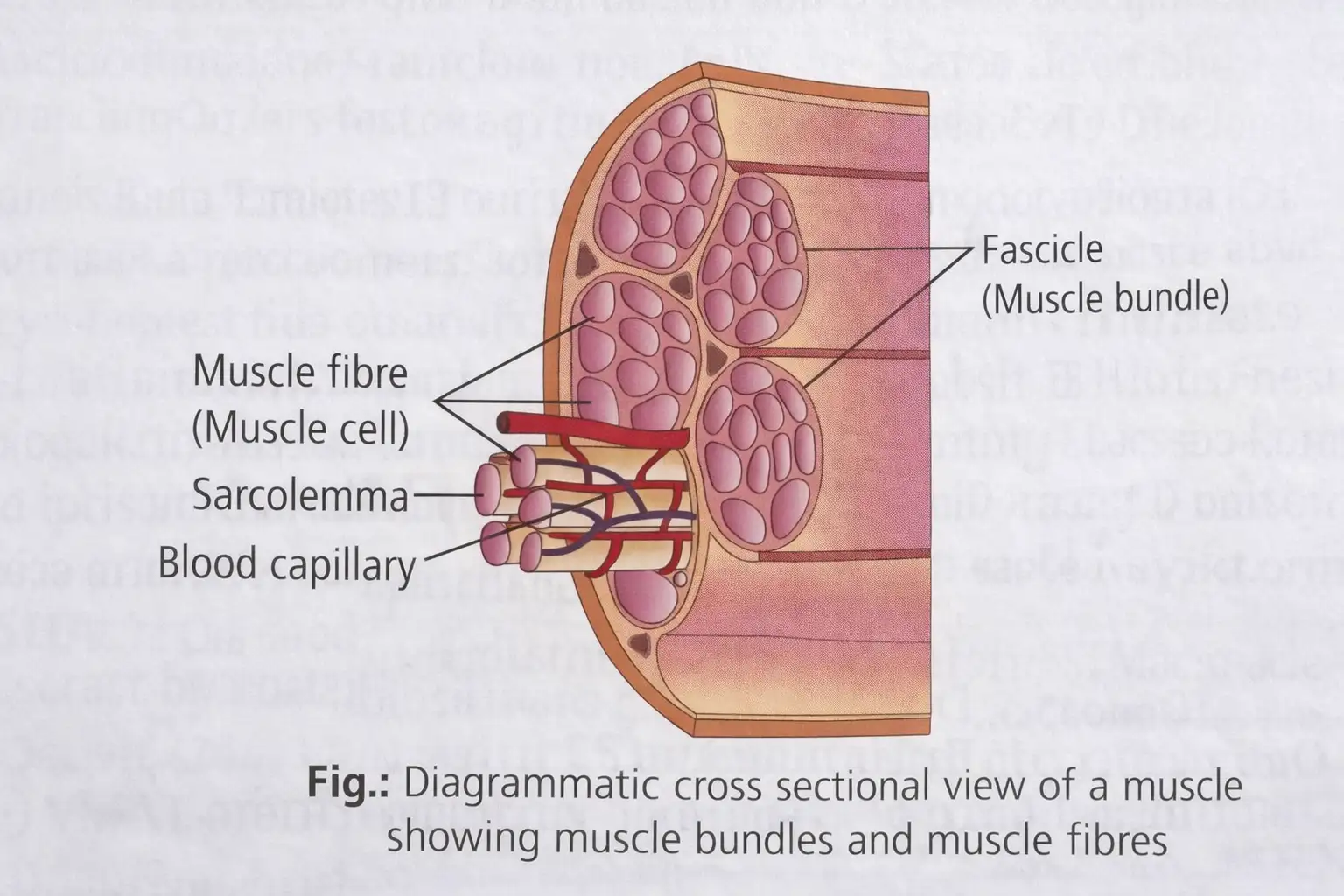
Myofibrils and sarcomeres
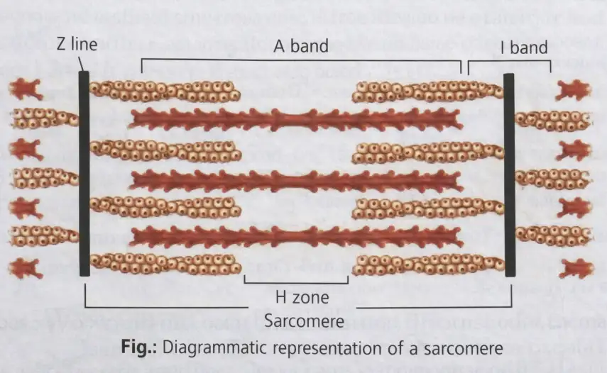
- Striations: result from the arrangement of thin actin and thick myosin filaments.
- I-band: light, isotropic band containing actin; its centre is bisected by the elastic Z-line (Z-disc/Krause’s membrane), where thin filaments attach.
- A-band: dark, anisotropic band containing myosin; thick filaments are joined at its centre by the M-line.
- Sarcomere: region between two successive Z-lines; it is the functional contractile unit of a myofibril and contains thick and thin filaments.
- H-zone: in a resting sarcomere, the central part of the thick filaments that is not overlapped by thin filaments.
Contractile Proteins
| Protein | Structure and role |
|---|---|
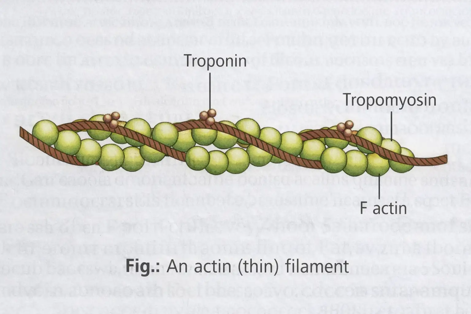
| Actin | Thin-filament backbone; two helically wound F-actin strands form the filament. Each F-actin is a polymer of G-actin monomers. |
| Tropomyosin | A double-stranded, alpha-helical rod attached to F-actin. At rest, it lies over actin’s active sites and prevents actin–myosin interaction. |
| Troponin | Complex protein present at regular intervals on tropomyosin. At rest, a subunit masks the myosin-binding sites on actin. |
| Myosin | Main component of the thick filament; its projecting heads form cross-bridges with actin. |
Myosin structure
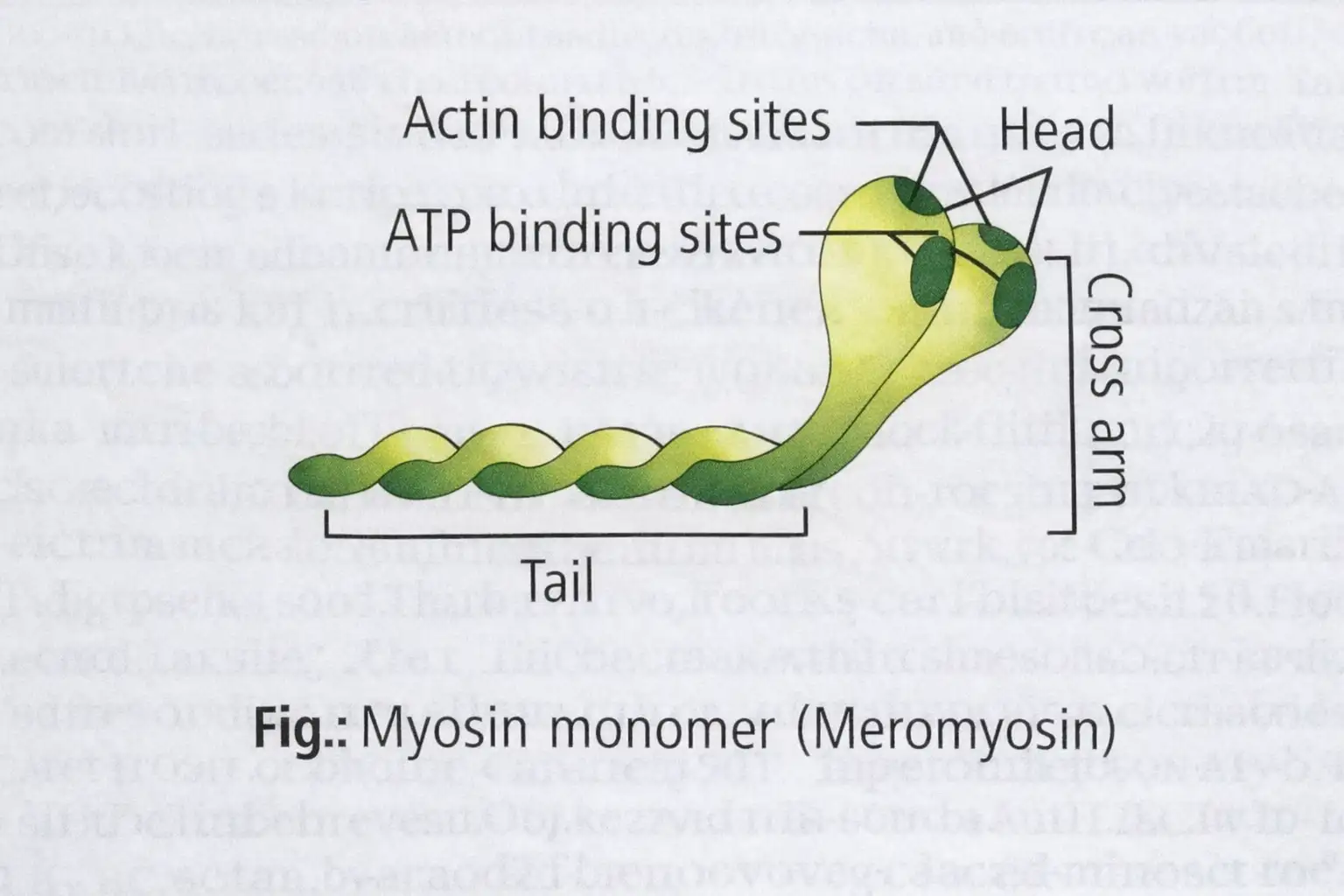
- Composition: muscle fibres contain four contractile proteins—myosin, actin, troponin and tropomyosin. A myosin molecule has two heavy polypeptide chains and four smaller light chains.
- Shape: two globular heads (containing heavy and light chains) and a long tail formed by the two twisted heavy chains.
- Head: contains actin-binding and ATP-binding sites and forms the cross-bridge.
- Trypsin digestion: splits myosin into heavy meromyosin (HMM) and light meromyosin (LMM); the globular HMM subfragment contains the binding sites.
Mechanism of Muscle Contraction
Sliding filament theory — Muscle contraction occurs when thin filaments slide over thick filaments, shortening the sarcomere.
Key points
- Proposed in 1954 by: A. F. Huxley and Ralph Niedergerke; and H. E. Huxley and Jean Hanson.
- Motor unit: a motor neuron together with the muscle fibres connected to it.
- Neuromuscular junction (motor end plate): junction between a motor neuron and the sarcolemma of a muscle fibre.
Contraction and relaxation sequence
- A CNS signal travels through a motor neuron to the neuromuscular junction; acetylcholine is released.
- Acetylcholine generates an action potential in the sarcolemma; it spreads through the fibre and causes calcium release into the sarcoplasm.
- Calcium binds to a troponin subunit, removing the masking of actin’s myosin-binding sites.
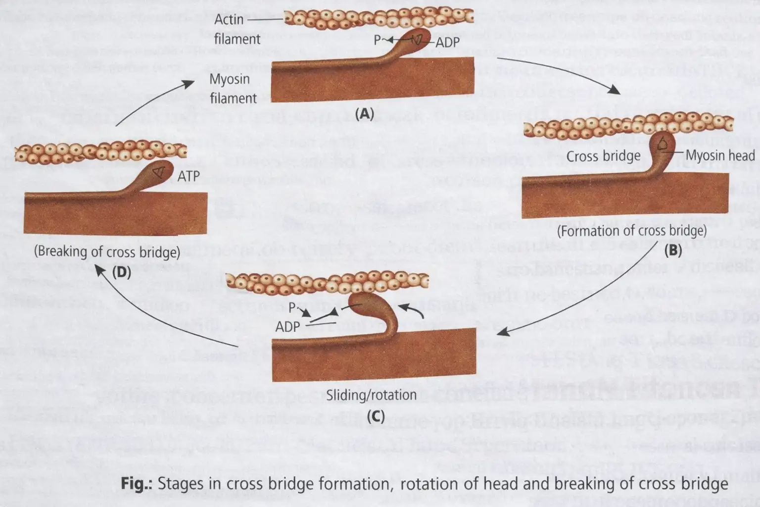
- ATP hydrolysis provides energy for the myosin head to bind actin, forming a cross-bridge.
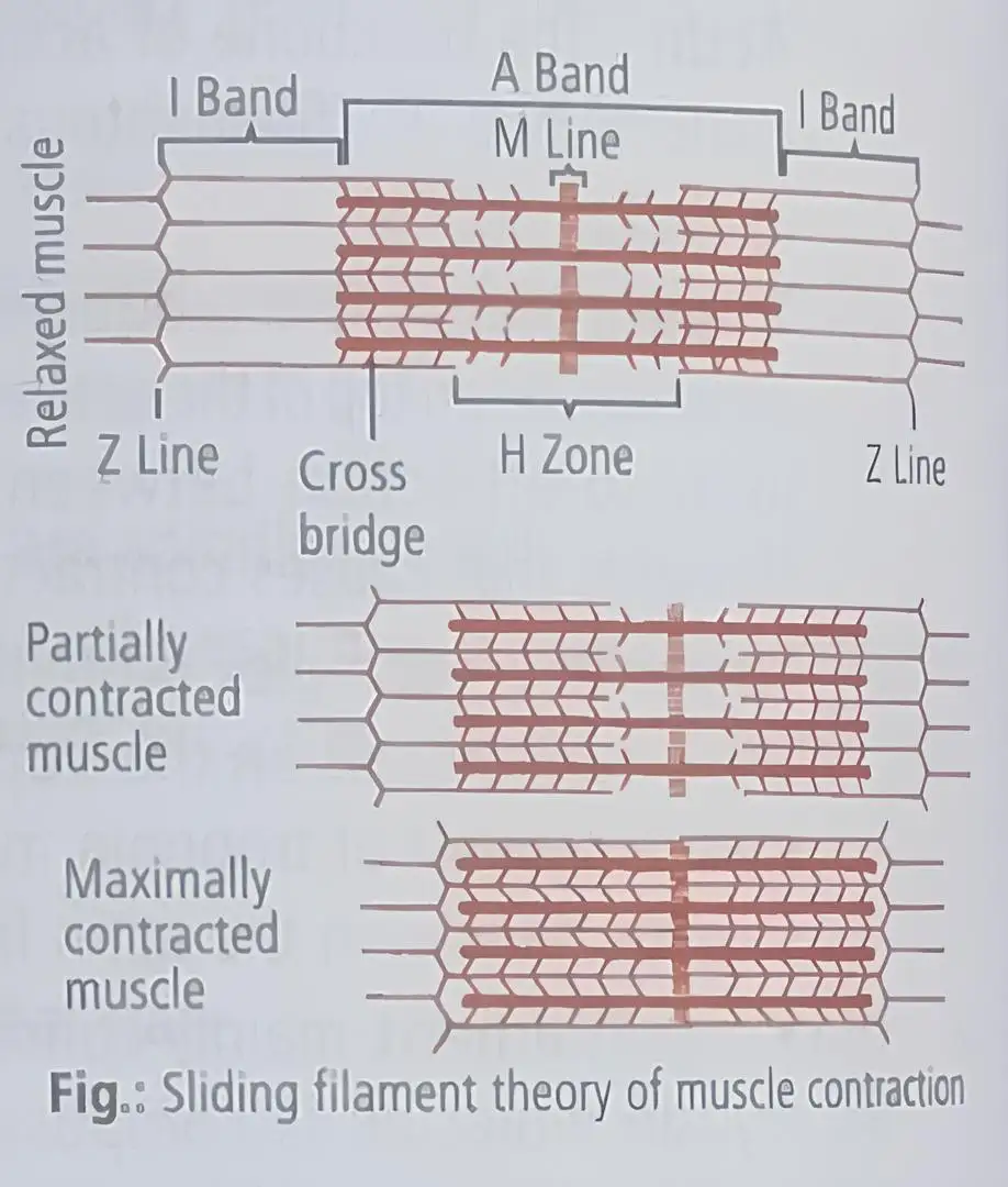
- The myosin head pivots and pulls actin towards the centre of the A-band; Z-lines move inward and the sarcomere shortens.
- Myosin releases ADP and inorganic phosphate, then binds a new ATP molecule; this breaks the cross-bridge.
- ATP is hydrolysed again and the cross-bridge cycle repeats, producing further filament sliding.
- When calcium is pumped back into the sarcoplasmic cisternae, actin sites are masked again; Z-lines return towards their original positions and the muscle relaxes.
| Feature | During contraction |
|---|---|
| Thin filaments | Slide towards the centre of the A-band. |
| Z-lines | Move towards each other, shortening the sarcomere. |
| I-band | Becomes narrower. |
| A-band | Retains its length. |
Skeletal Muscle Fibres and Related Concepts
| Feature | Red fibres | White fibres |
|---|---|---|
| Myoglobin and colour | High myoglobin; red | Low myoglobin; pale or whitish |
| Mitochondria | More numerous | Less numerous |
| Sarcoplasmic reticulum | Less abundant | More abundant |
| Energy and fatigue | Mainly aerobic oxidation; little lactic acid accumulation | Depend more on anaerobic oxidation; considerable lactic acid accumulates during strenuous work |
| Contraction | Slow; can work for longer periods | Fast; work for shorter periods |
| Example | Extensor muscles of the back | Eyeball muscles |
All-or-none law — A muscle fibre contracts maximally once it is activated; it does not contract by a partial amount.
Oxygen debt, fatigue and muscle pain
- Vigorous exercise can reduce oxygen supply to muscle.
- Muscle then breaks down glycogen anaerobically; pyruvic acid is converted to lactic acid.
- Lactic acid accumulation contributes to muscle fatigue and pain.
- Extra oxygen is needed afterwards to oxidise the accumulated lactic acid aerobically; this is the muscle’s oxygen debt.
Rigor mortis — Stiffening of the body after death because ATP is unavailable to detach actin and myosin; it persists until protein decomposition begins.
Antagonistic muscles — Muscles that produce opposite actions at a joint.
| Muscle | Action | Functional name |
|---|---|---|
| Biceps | Bends or flexes the arm | Flexor |
| Triceps | Straightens or extends the arm | Extensor |
What happens to the A-band and I-band during muscle contraction?
How does calcium initiate muscle contraction?
Why does rigor mortis occur?
Quick recap
- Muscles are mesodermal tissues with excitability, contractility, extensibility and elasticity; the three types are skeletal, cardiac and smooth.
- Skeletal muscle fibres are long, cylindrical and multinucleate; their parallel myofilaments form myofibrils, and the sarcomere between two Z-lines is the contractile unit.
- The I-band is isotropic and contains actin; the A-band is anisotropic and contains myosin.
- Troponin and tropomyosin regulate actin’s binding sites; calcium exposes these sites and ATP-powered myosin cross-bridges slide actin inward.
- During contraction, the I-band narrows while the A-band retains its length; calcium removal allows relaxation.
- Red fibres contract slowly for longer, while white fibres contract quickly for shorter periods; vigorous exercise may cause lactic acid accumulation and oxygen debt.
- ATP shortage causes rigor mortis, while biceps and triceps illustrate opposing flexor and extensor actions.
Skeletal System
Overview and Types of Skeleton
Axial Skeleton
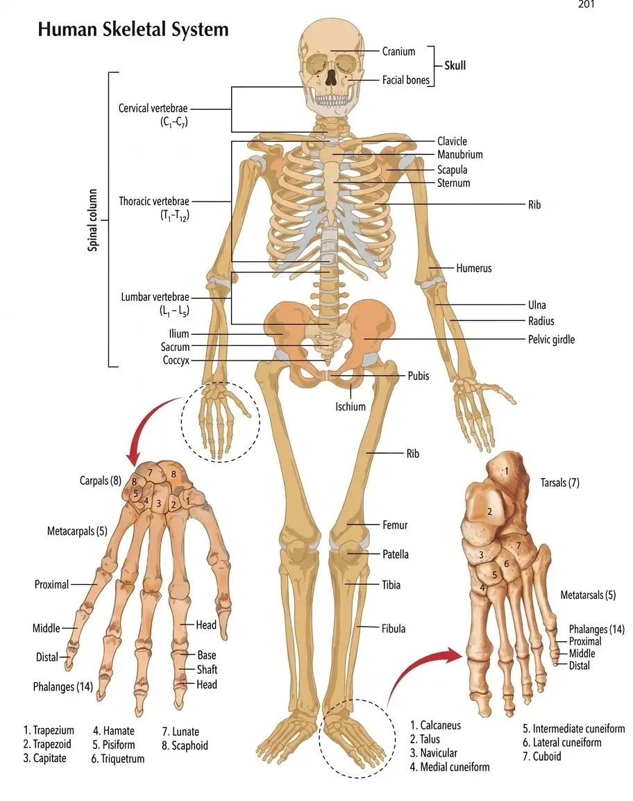
Axial skeleton — The 80 bones lying along the body’s longitudinal axis support and protect the head, neck and trunk.
Skull
Skull bones and articulations
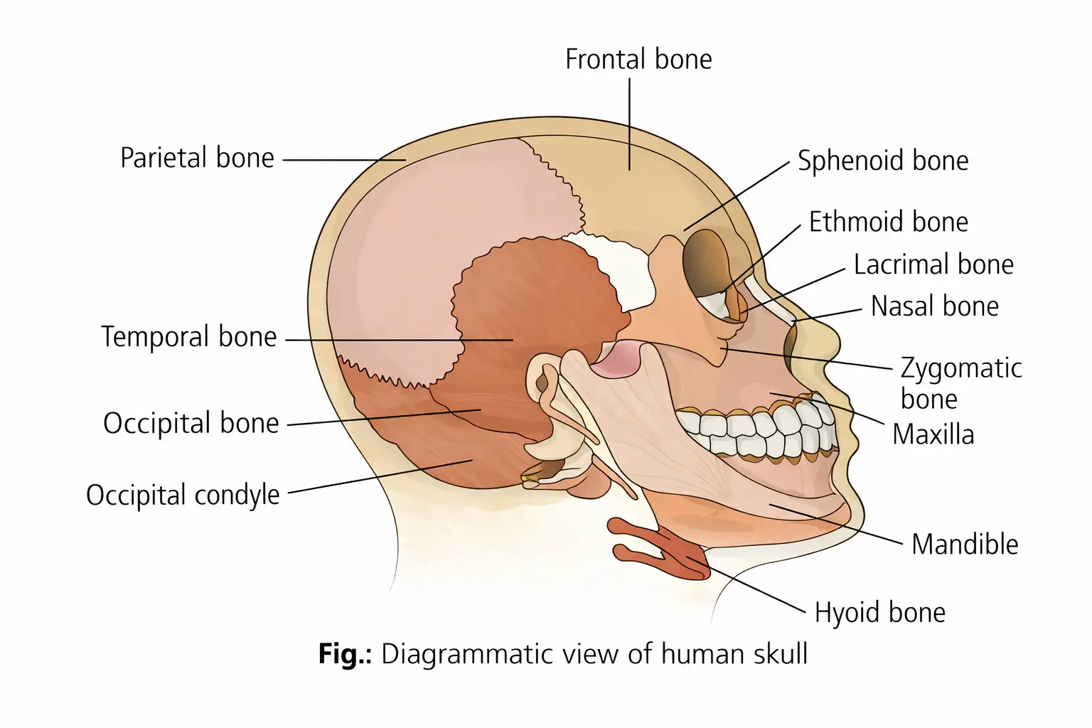
- Total: 22 bones—8 cranial bones and 14 facial bones.
- Cranial bones form the hard cranium that protects the brain; facial bones form the front part of the skull.
- Hyoid: a single U-shaped bone at the base of the buccal cavity.
- Ear ossicles: each middle ear contains three tiny bones—malleus, incus and stapes.
- Dicondylic skull: two occipital condyles articulate with the superior end of the vertebral column.
Vertebral Column
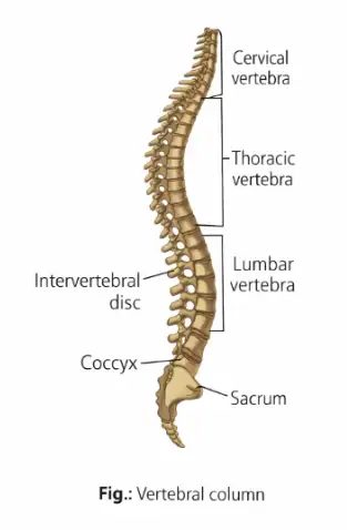
The formula shows the vertebral regions and their counts, including the fused sacral and coccygeal units.
Sternum and Ribs
Appendicular Skeleton
Limb bones: key features
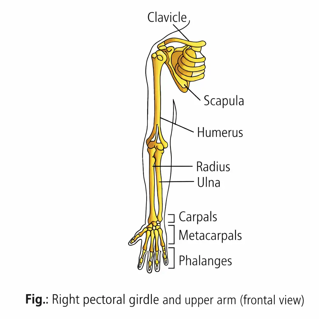
- Carpals are wrist bones; metacarpals form the palm; phalanges form the digits.
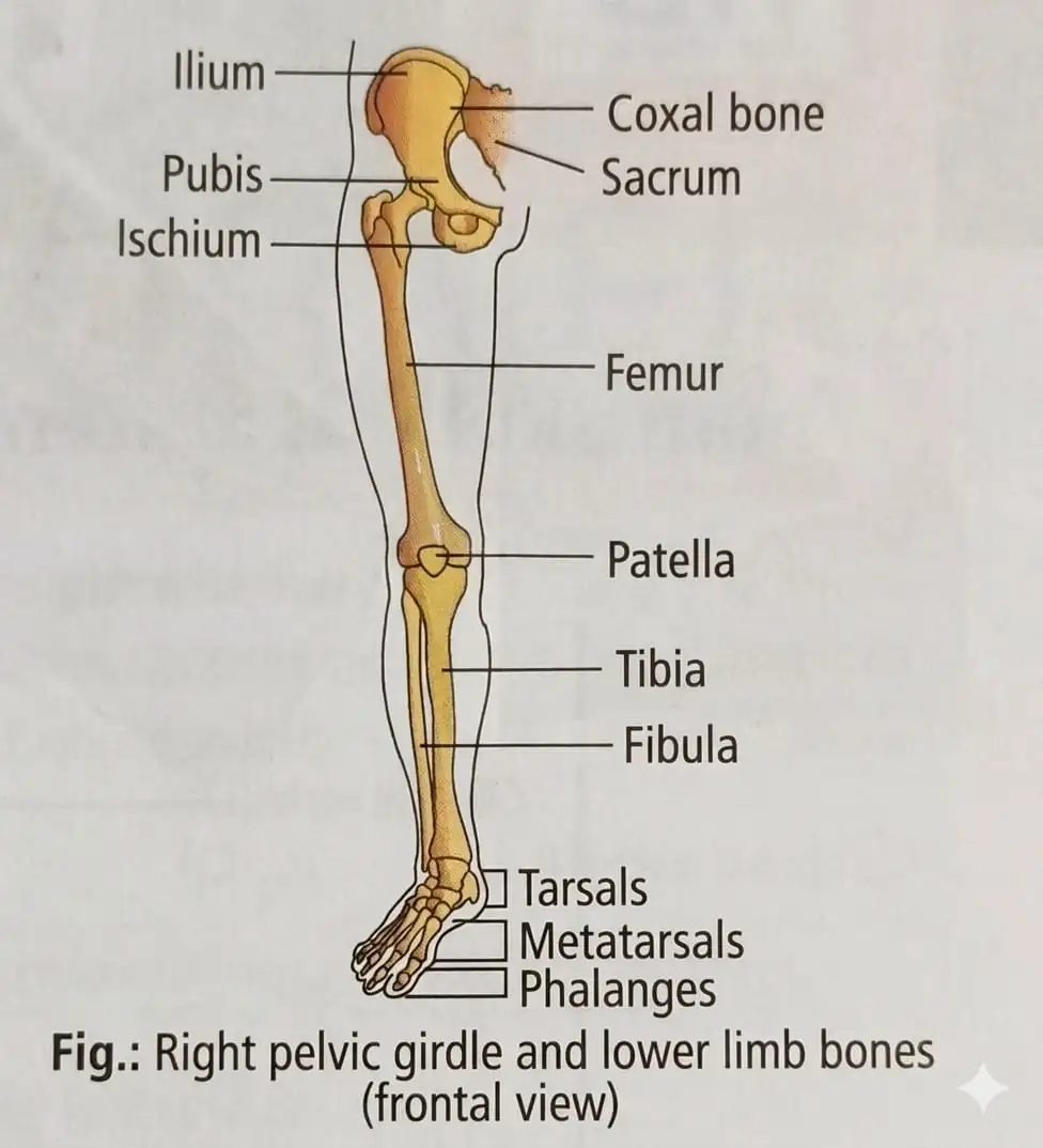
- Tarsals are ankle bones; metatarsals form the foot.
- Patella is a cup-shaped bone covering the knee ventrally; it is also called the kneecap.
- Femur is the longest, largest and strongest bone in the body.
- Lower limbs support body weight and help with propulsion.
Pectoral Girdle
Pelvic Girdle
Name the vertebral regions and give their counts.
Classify the 12 pairs of ribs.
What forms the shoulder joint and the hip joint?
Quick recap
- The adult human skeleton has 206 bones: 80 axial and 126 appendicular.
- The skull has 22 bones; the hyoid and middle-ear ossicles are additional axial bones.
- The vertebral column has 26 units across five regions; atlas is the first cervical vertebra and axis is the second.
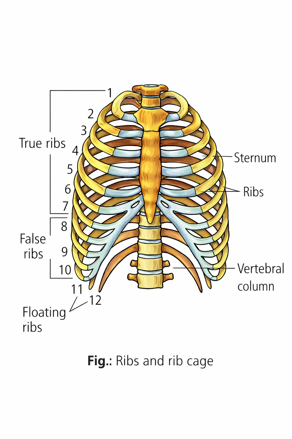
- There are 12 pairs of ribs: 7 true, 3 false and 2 floating.
- Each limb has 30 bones; the femur is the body’s longest, largest and strongest bone.
- The pectoral girdle connects upper limbs; the pelvic girdle connects lower limbs to the axial skeleton.
Joints
Joints and Movement
Joint — A joint is a point of contact between bones, or between a bone and cartilage.
How joints enable movement
- Role: Joints are essential for movements involving the bony parts of the body.
- Movement: Muscles generate force that acts through joints; the joint functions as a fulcrum.
- Movability: The degree of movement differs among joints.
- Ligaments and tendons: Ligaments join bones; tendons connect muscles to bones.
Structural Classification of Joints
| Type | Structure and mobility | Examples |
|---|---|---|
| Fibrous | Bones are joined by dense fibrous connective tissue; no movement. | Skull sutures; joints between teeth and the maxilla or mandible. |
| Cartilaginous | Bones are joined by cartilage; movement is limited. | Adjacent vertebrae; pubic symphysis. |
| Synovial | A fluid-filled synovial cavity lies between articulating bone surfaces; considerable movement is possible. | Ball-and-socket, hinge, pivot, gliding and saddle joints. |
Synovial Joint Types and Examples
- Ball-and-socket: Humerus and pectoral girdle.
- Hinge: Knee joint.
- Pivot: Atlas and axis.
- Gliding: Between carpals.
- Saddle: Between the carpal and metacarpal bones of the thumb.
Name the three structural types of joints and state the mobility of each.
Quick recap
- Joints connect bones to bones or bones to cartilage and enable movement.
- Muscle force acts through a joint, which functions as a fulcrum.
- Ligaments join bones; tendons connect muscles to bones.
- Fibrous joints are immovable; examples include skull sutures and tooth sockets.
- Cartilaginous joints permit limited movement; examples include vertebrae and pubic symphysis.
- Synovial joints have a fluid-filled cavity and allow considerable movement.
- Synovial types include ball-and-socket, hinge, pivot, gliding and saddle joints.
Disorders of Muscular and Skeletal System
Skeletal System Disorders
Osteoporosis — An age-related disease in which bones lose minerals and fibres from their matrix.
Muscular System Disorders
Muscular dystrophy — A disorder caused by a mutated gene on the X chromosome that fails to produce dystrophin in skeletal muscles.
Myasthenia gravis — An autoimmune disorder affecting the neuromuscular junction and causing fatigue, weakening and paralysis of skeletal muscles.
Bone Mineralisation and Joint Disorders
Osteomalacia or rickets — A childhood disorder in which bones contain insufficient calcium and phosphorus; the source uses osteomalacia and rickets as alternative names.
Arthritis — Inflammation of the joints.
Gout — A disorder caused by defective purine metabolism, leading to excess uric acid and its salts accumulating in joints.
Tetany — A condition involving rapid muscle spasms caused by low calcium levels in body fluid.
How does dystrophin deficiency affect muscle contraction in muscular dystrophy?
Name the three types of arthritis listed in this lecture.
What causes gout and tetany?
Quick recap
- Osteoporosis is age-related bone-matrix loss associated with hormonal imbalance or calcium and vitamin D deficiency.
- Muscular dystrophy involves an X-chromosome mutation, deficient dystrophin and failure of muscle contraction.
- Myasthenia gravis is an autoimmune disorder of the neuromuscular junction.
- Osteomalacia, also called rickets in the source, occurs in childhood when bones lack sufficient calcium and phosphorus.
- Arthritis is joint inflammation; listed types are rheumatoid, osteoarthritis and infectious arthritis.
- Gout is linked to excess uric acid in joints; tetany is linked to low body-fluid calcium.
NEET Zoology — ALL CHAPTERS
1
Structural Organisation in Animals
2
Breathing and Exchange of Gases
3
Body Fluids and Circulation
4
Excretory Products and their Elimination
5
Locomotion and Movement
6
Neural Control and Coordination
7
Human Reproduction
8
Reproductive Health
9
Evolution
10
Microbes in Human Welfare
11
Organism and Population
12
Ecosystem
13
Biodiversity and Conservation